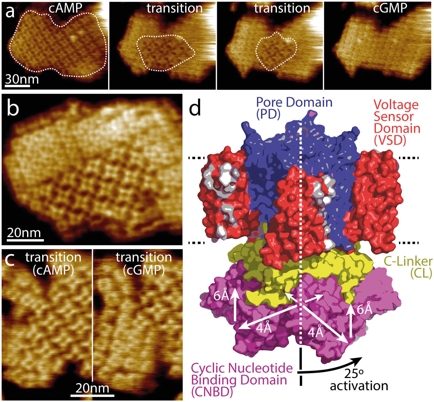
Figure 2. Dynamics of ligand-induced conformational changes in SthK by real-time HS-AFM imaging.

(a) HS-AFM time-lapse high-resolution image sequence of a SthK 2D-crystal initially in 0.1mM cAMP and exposing CNBDs. Upon addition of 7mM cGMP, SthK channels undergo a conformational change progressively from the borders to the center of the membrane patch (dotted outline). (b) High-resolution topography of a membrane containing well-ordered channels in both conformations. (c) High-resolution topographs during a cAMP to cGMP transition where the majority of the molecules are in the cAMP (left) and in the cGMP (right) conformation, respectively. (d) Model of SthK in the activated state: Upon activation, the CNBDs rotate by ~25° clockwise (when viewed from the intracellular side) and move by ~6 Å towards the membrane and by ~4 Å outwards from the four-fold axis (note, this activated state illustration is a cartoon using domains of the SthK structure repositioned according to the displacements found by HS-AFM).
