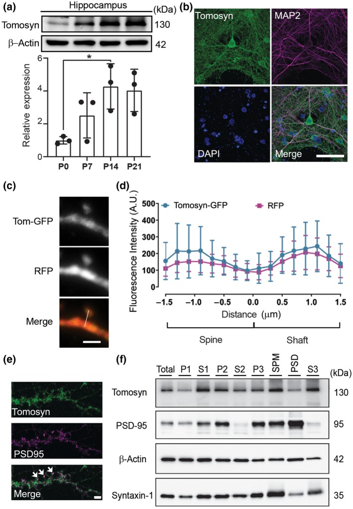
FIGURE 1.

Tomosyn is localized in both pre and postsynaptic compartments. (a) Western blot analysis shows the developmental expression pattern of tomosyn protein in the hippocampus at P0, P7, P14, and P21; n = 3. (b) Confocal immunofluorescence images showing tomosyn (green), MAP2 (red), and DAPI (blue) from a cultured hippocampal neuron at 15 DIV. Scale bar, 50 μm. (c) Confocal images of a dendritic spine of a cultured neuron transfected with RFP and tomosyn‐GFP. Scale bar, 2 µm. (d) Fluorescence intensity of tomosyn‐GFP and RFP in the dendritic spine versus the dendritic shaft was quantified by line scan (shown in c); n = 29. (e) Confocal images of a dendritic segment of a cultured neuron immunostained with tomosyn and PSD‐95 antibodies. Arrows indicate the colocalization of tomosyn and PSD‐95. Scale bar, 2 μm. (f) Synaptic fractionation of forebrain lysates (total) shows the subcellular localization of tomosyn in P21 mouse brains. P1, nuclei; S1, cytosol/membranes; P2, crude synaptosomes; S2, cytosol/light membranes; P3, synaptosomes; SPM, synaptic plasma membranes; PSD, postsynaptic density; S3, synaptic vesicles. Brightness and contrast were adjusted in b and e for better visualization
