FIGURE 6.

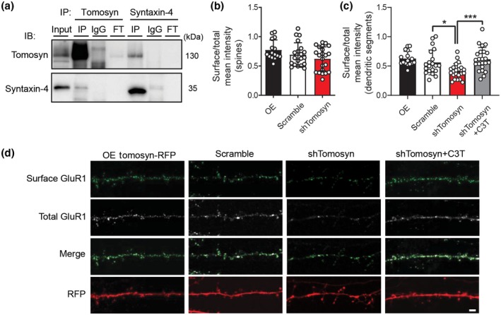
Surface expression of GluR1 subunits is reduced in tomosyn knockdown neurons. (a) Western blot analysis of co‐immunoprecipitation experiments in cultured cortical neurons. Cultured cortical neuron lysates at 15 DIV were immunoprecipitated with either anti‐tomosyn antibody, rabbit IgG control, anti‐syntaxin‐4 antibody, or mouse IgG control. Immunoblot was probed with tomosyn and syntaxin‐4 antibodies. FT = flow through. Quantification of the surface expression of pHluorin‐GluR1 in (b) dendritic spines and (c) dendritic segments. Cultured neurons were co‐transfected with either Tomosyn‐RFP (OE), scrambled shRNA, or shTomosyn and pHluorin‐GluR1. Tomosyn knockdown neurons were treated with the RhoA inhibitor, C3T. *p < 0.05, ***p < 0.001, one‐way ANOVA with Dunnett's multiple comparisons test. n = 16 OE, 20 scramble, 22 shTomosyn, and 23 shTomosyn + C3T. (d) Representative confocal images showing surface staining for pHluorin‐GluR1 in cultured hippocampal neurons at 15 DIV. Scale bar, 2 µm
