Figure 1.

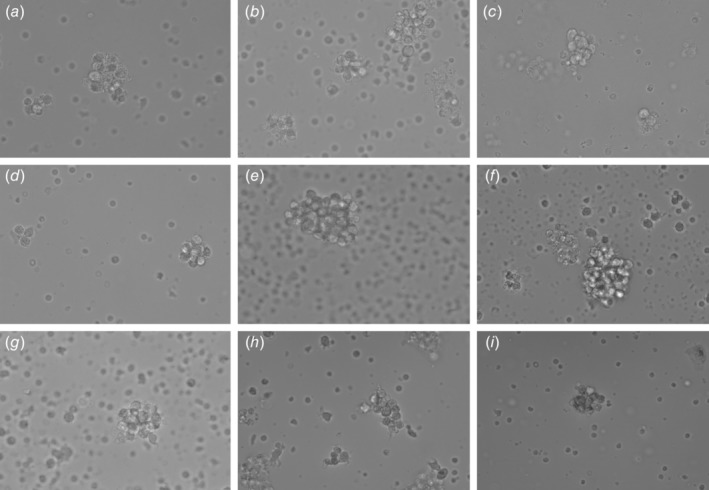
Cell assemblages on Day 5. Viable intact cell assemblages (white arrow) were imaged under a phase contrast microscope at 40× magnification. Samples from various cancer types are depicted (a) breast, (b) lung, (c) prostate, (d) stomach, (e) gallbladder, (f) kidney, (g) bladder, (h) buccal mucosa and (i) pancreas. Field width is ~160 μm.
