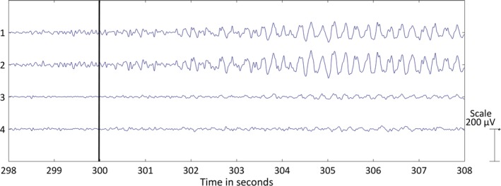
FIGURE 2.

Left temporal lobe seizure recorded with behind‐the‐ear electroencephalographic (EEG) setup. The four bipolar EEG channels are shown over a period of 10 seconds: (1) crosshead 1, (2) crosshead 2, (3) unilateral left, (4) unilateral right. The bipolar EEG channels were filtered with a bandpass filter (1‐25 Hz). The black horizontal line at 300 seconds depicts the seizure onset
