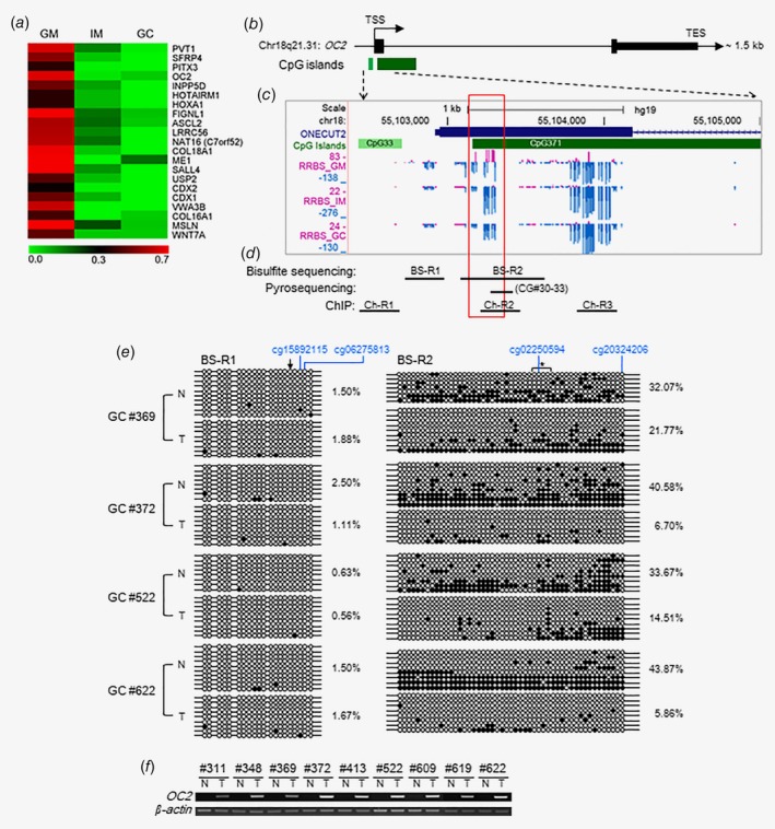
Figure 1.

Methylation profile of early‐onset hypomethylated DMPs and methylation signatures at promoter‐proximal DNA of OC2. (a) Heat map of selected early‐onset hypomethylated DMPs showing relative frequencies of methylated reads to total reads around gene promoters in GM, IM and GC cells from RRBS analysis. (b) Gene structure of OC2 on human chromosome 18q21.31. Map was modified from the UCSC Genome Browser (http://genome.ucsc.edu/). The distance from TSS to TES is ~1.5 kb. TSS, transcription start site; TES, transcription end site; thick black bars, exons; thin black bars, 5′‐ or 3′‐untranslated regions; green bars, CpC islands containing 33 and 371 CpGs, respectively. (c) RRBS methylome profiles around the enlarged OC2 exon1 DNA in paired GM, IM and GC cells from mirroring UCSC Genome Browser (hg19). Vertical lines indicate methylation scores of individual CpGs: Methylation and unmethylation scores are displayed as purple upward and blue downward bars. Red rectangle highlights differentially methylated region in GM compared to IM or GC. (d) Strategy for analysis in our study. Bisulfite sequencing was performed at promoter region BS‐R1 (20 CpGs, −117–41 nt from TSS) and the promoter‐proximal region BS‐R2 (49 CpGs, 209–685 nt). Pyrosequencing was performed at four CpGs (#30–33) between 418 and 448. ChIP was performed at Ch‐R1 (−451 to −270 nt), Ch‐R2 (356–541 nt), and Ch‐R3 (1,014–1,194 nt). (e) Bisulfite sequencing was performed with four paired GC tissues (#369, 372, 522 and 622; N, normal; T, tumor). Black and white circles indicate methylated and unmethylated CpG sites, respectively. Each row represents a single clone. Numbers on the right represent mean percentage of CpG sites that were methylated in each sample tissue. Four CpG sites (blue colored) in top indicate CpG positions examined in Figures 2 a and 2b. Arrow in top left indicates TSS. Asterisk in top right indicates four CpG sites used for pyrosequencing. (f) RT‐PCR in clinical tissues. OC2 expression was examined in nine paired gastric tumor tissues, including the four paired tissues used for bisulfite sequencing. β‐Actin was used as an internal control.
