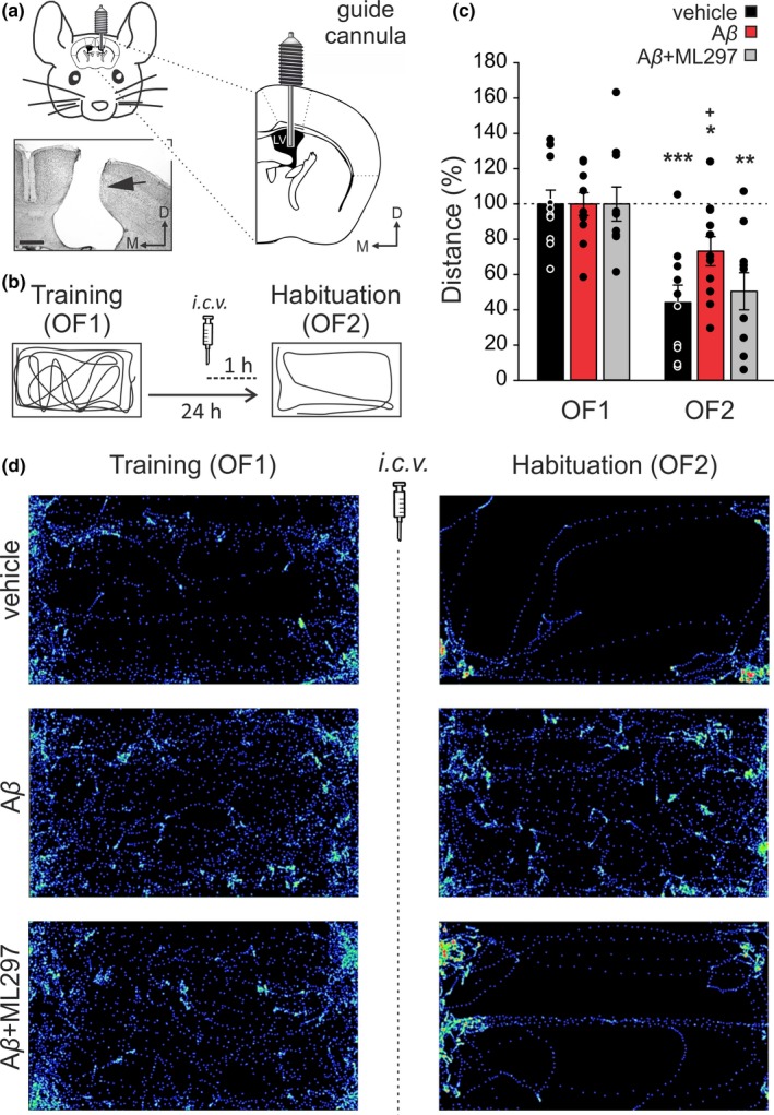
Figure 4.

Effects of G‐protein‐gated inwardly rectifying potassium channels (GirK) activation on mice intracerebroventricular (i.c.v.)‐injected with amyloid‐β (Aβ)1–42 during hippocampal‐dependent habituation to an open field. (a, b) Experimental design. (a) Pictures illustrate how mice were prepared for drug administration. A stainless‐steel guide cannula was implanted chronically on the left ventricle [1 mm lateral, 0.5 mm posterior to bregma, and 1.8 mm from the brain surface]. The photomicrograph serves as histological verification of cannula position (black arrow). Scale Bar 500 µm. LV, Lateral Ventricle; D, dorsal; M, medial. (b) Open field habituation test. Mice were exposed for 15 min to the same open field (OF) two consecutive times with a 24‐hr interval (OF1, Training trial, and OF2, Habituation or retention trial). Habituation memory levels were determined by measuring exploration behavior. I.c.v. injections were performed 1 hr before OF2. For each mouse, traveled distance was automatically tracked and recorded using a LABORAS® system (Metris, Hoofddorp, The Netherlands) on the basis of detecting vibrations of the movements of each animal. Diagrams represent an example of the path followed by a control (vehicle‐injected) animal during both training (OF1) and habituation (OF2) sessions. (c) Total horizontal distance traveled during the 15‐minsession for vehicle (n = 10 animals), Aβ1–42 (n = 11) and Aβ1–42 + ML297 (n = 10) ‐treated mice before (OF1) and after (OF2) drug administration. Data were normalized as percentage of the movement in the training (OF1) session. Differences between OF1 and OF2 are indicated by asterisks (*p < .05; **p < .01; ***p < .001). Differences versus. control (vehicle) within OF2 session are indicated by crosses (+, p < .05). (d) LABORAS®‐generated images illustrate the distance and path traveled by a representative animal of each experimental group during OF1 and OF2 sessions
