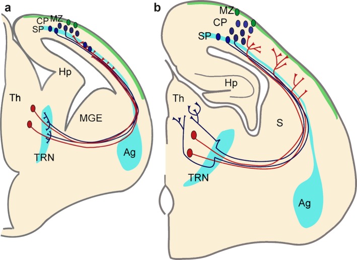
Figure 1.

Functional correlation between the developing thalamocortical projections, cortical SP zone and thalamic reticular nucleus. (a) Corticofugal (blue) and thalamocortical (red) axons extend towards each other at early stages during embryonic development and reach close to their targets. However, they both stop short of their ultimate targets and corticofugal projections from subplate and layer VI accumulate in the thalamic reticular nucleus (TRN) and thalamocortical projections in subplate, respectively. Both compartments contain largely transient cells that get integrated into circuits during these early stages. (b) Towards the middle of the first postnatal week corticofugal and corticopetal axons enter the thalamus (Th) and cortical plate (CP), respectively, where they arborize and establish their contacts with their ultimate targets in thalamus and neocortex. There are signs of fibre decussations in the TRN and in the subplate indicating some rearrangements during development. Pale blue areas (amygdala, subplate and thalamic reticular nucleus) represent brain regions sharing gene expression patterns. Ag: amygdala; Hp: hippocampus; MGE: medial ganglionic eminence; MZ: marginal zone (green area); S: striatum. Figure from Montiel et al. (2011).
