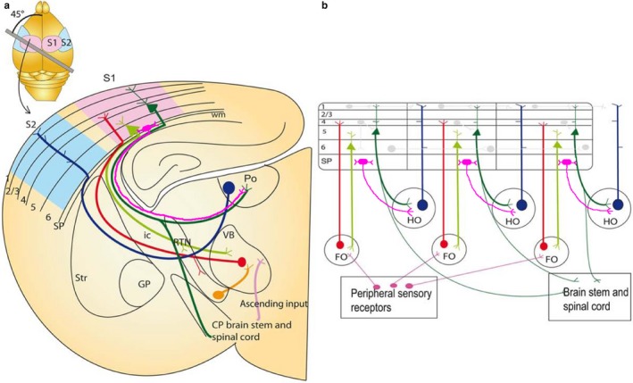
Figure 2.

Thalamocortical circuits in the adult on an idealized section containing somatosensory cortical connections (a) and schematic representation of the two major sets of thalamic projection neurons (b). (a) Inset: outline of the mouse brain with the line indicating the plane of section to obtain thalamocortical slice containing S1 with intact thalamocortical projections. For clarity, S2 cortex connectivity is also indicated in the idealized section, although a different plane of section would be required to maintain connections. Main image: coronal schematic demonstrating the specificity of the connections between the cortex and thalamus using the somatosensory system as an example. The first order VB thalamic nucleus receives somatosensory peripheral input (pink). The VB then projects axons (red) to layer 4 of the primary somatosensory cortex (S1; light blue). Layer 6 “modulator” neurons (light green) in S1 project back to the VB. Layer 5 neurons (dark green) in S1 project to subcerebral structures and make a collateral branch to a higher order thalamic nucleus, for example, posterior thalamic nucleus (Po). Some of the persistent subplate cells at the bottom of layer 6 (SP, bright pink) selectively project to higher order thalamic nucleus Po, without giving a collateral to thalamic reticular nucleus (RTN). The higher order nuclei then project (dark blue) to an area of cortex that is different from the one they received input from (for example S2; light pink). This projection pattern generates an open loop. (b) Schematic illustration of the possible functional circuits generated by this reoccurring open loop connectivity and how persistent subplate (SP) can regulate the transthalamic cortico‐cortical communication through their projections (bright pink) to higher order thalamic nuclei. Sensory information is relayed through the first order thalamic nucleus to the cortex (red). This cortical area then projects from layer 6 reciprocally back to the first order nucleus (light green). Each area is also non‐reciprocally connected to a higher order thalamic nucleus. The layer 5 input to the thalamus (dark green) is an “efference copy” of the layer 5 output to the motor system in the brainstem and spinal cord. This copy is forwarded to a higher cortical area (blue). Some persistent subplate (SP) in layer 6b selectively project to higher order thalamic nuclei and in a position to regulate the transthalamic cortico‐cortical communication. Direct cortico‐cortical connections are also depicted between cortical layers and cortical areas (pale grey lines). These circuits enable cortical areas to act with other cortical areas and motor apparatus in a coordinated manner. CP: cerebral peduncle; FO: first order thalamic nuclei; GP: globus pallidus; HO: higher order thalamic nuclei; ic: internal capsule; RTN: reticular thalamic nuclei; SP: subplate; Str: striatum; S1: primary somatosensory cortex; S2: secondary association somatosensory cortex; Po: posterior thalamic nuclei; VB: ventrobasal thalamic nuclei; wm: white matter. Figure is modified from Grant et al. (2012) that was inspired by Guillery and Sherman (2002) and modified based on the results of Hoerder‐Suabedissen, Hayashi et al., 2018.
