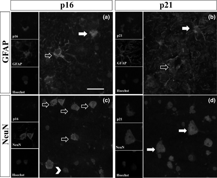
Figure 2.

Representative images of double immunofluorescence for p16 and p21 with GFAP or NeuN in the FACx of ALS/MND donors. (A) Glial p16 (red) co‐localized with processes of GFAP + astrocytes (green) (arrows), but not all GFAP expressing astrocytes were positively labelled for p16 (arrow outlines). (B) Glial p21 (red) co‐localized with GFAP + astrocytes (green) (arrows); there was also a population of GFAP + astrocytes that did not express p21 (arrow outlines). (C) Expression of p16 (red) was only localized to glial cells (arrowheads) but not to neurones (arrow outlines). (D). Co‐localization of p21 and NeuN confirmed expression of p21 in neurones (arrows). Scale bar represents 25 μm. ALS/MND, Amyotrophic lateral sclerosis/motor neurone disease; GFAP, glial fibrillary acidic protein; FACx, frontal association cortex.
