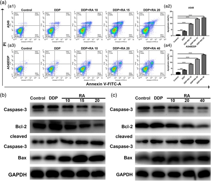
Figure 3.

Induction of cell apoptosis by RA in NSCLC cell lines. (a1–a4) Induction of apoptosis by 1.5 or 15 μg/ml DDP and DDP combined with various concentrations of RA in A549 (a1, a2) and A549DDP (a3, a4) cells evaluated by Annexin‐V‐FITC/PI staining. (b) Western blot analysis of Bcl‐2, Bax, Caspase‐3 and cleaved Caspase‐3. A549 cells were treated with DDP and combined with various concentrations of RA medication for 48 hr. GAPDH was used as the loading control. (c) Western blot analysis of Bcl‐2, Bax, Caspase‐3 and cleaved Caspase‐3. A549 cells were treated with DDP and combined with various concentrations of RA medication for 48 hr. GAPDH was used as the loading control. **p < 0.01; ***p < 0.001; versus control. DDP, cisplatin; NSCLC, non‐small cell lung cancer; PI, propidium iodide; RA, rosmarinic acid
