
Fig. 4 |. The microtubule cytoskeleton coordinates nuclear probing with cellular locomotion.
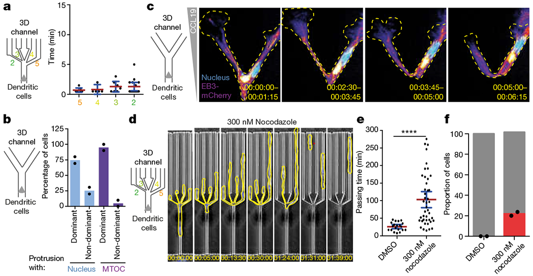
a, Retraction of cellular protrusions (from non-chosen pores) on passage of the MTOC into the chosen pore (three experiments, 34 cells). b, Path choice of DCs in Y-junction channels on translocation of the nucleus or the MTOC into one of the Y branches (two experiments, 46 cells). c, Microtubule plus ends, labelled with EB3–mCherry (fire colour-coded intensity), and nucleus (Hoechst stain, cyan) in a DC migrating through a Y-junction channel (time indicated; representative of three experiments). Images depict maximal intensity projections of 15 frames (5 s intervals). d, DCs (outlined in yellow) migrating through a decision point with pores 2, 3, 4 and 5 μm wide in the presence of 300 nM nocodazole (representative of two experiments). The red star highlights loss of cellular integrity. e, Quantification of the passing time in the experiments represented in d (two experiments, 63 cells; Mann–Whitney test; ****P < 0.0001). f, Quantification of the cellular integrity in the experiments represented in d (two experiments, 61 cells). Data in c and f are mean ± s.d.; data in a and e are mean ± 95% CI.