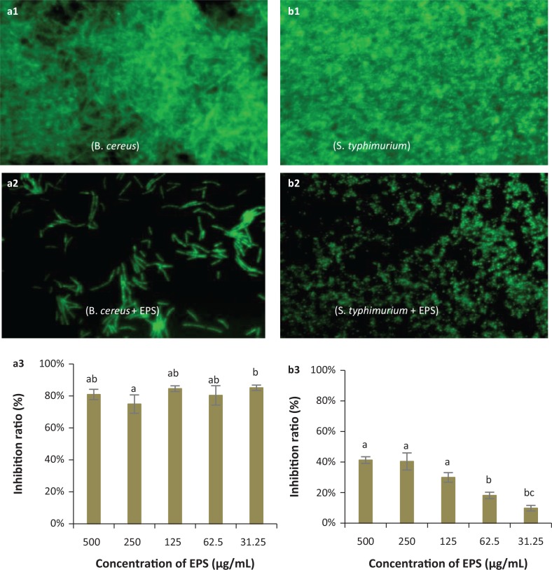
Fig. 6.
Representative micrographs (40×) showing the inhibitory effects of EPS on biofilm formation by pathogenic bacteria: Bacillus cereus (a1, a2, a3) and Salmonella typhimurium (b1, b2, b3) and spectrophotometric analyses of EPS biofilm inhibition. (a1, b1): Negative controls (pathogen only, no EPS) for biofilm formation by fluorescence microscopy; (a2, b2): Biofilm formation in the presence of EPS observed by fluorescence microscopy; (a3, b3): Inhibition ratios of EPS against B. cereus and S. typhimurium biofilms respectively. a,b,cDifferent letters indicate significant differences between concentrations (P < 0.05).

