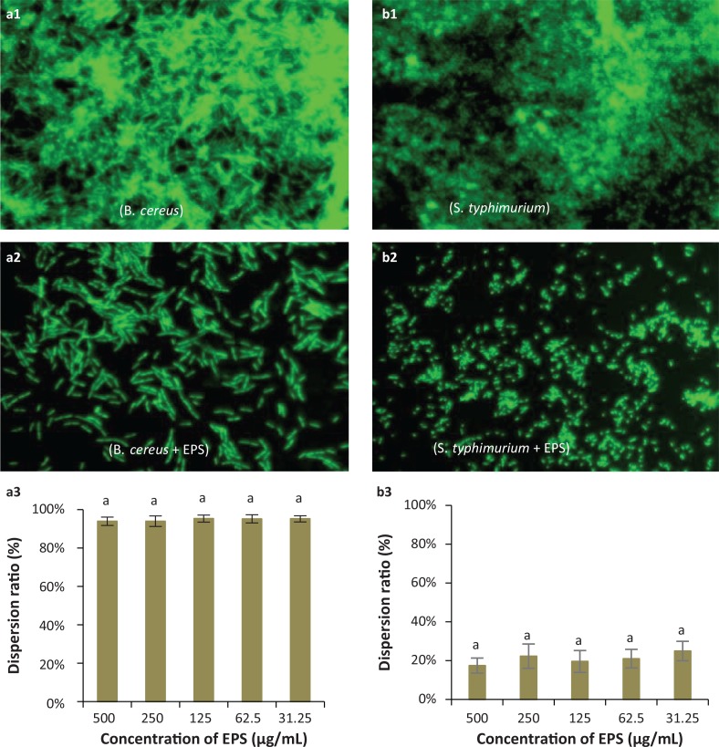
Fig. 7.
Representative micrographs (40×) showing the dispersion activity by EPS against established biofilms of the pathogenic bacteria Bacillus cereus (a1, a2, a3) and Salmonella typhimurium (b1, b2, b3), and spectrophotometric analyses of EPS-mediated dispersion of biofilms produced by these species. (a1, b1): Observation by fluorescence microscopy of negative controls (pathogens only, no EPS) for biofilm dispersion; (a2, b2): Observation by fluorescence microscopy of biofilm dispersion by Lactobacillus coryniformis NA-3-derived EPS; (a3, b3): Dispersion ratios of EPS on different bacterial biofilms. a,b,cDifferent letters indicate significant differences between treatments (P < 0.05).

