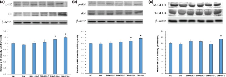
Fig. 5.
Expression of insulin-signaling-related proteins in the muscle. Protein expression of p-IR & IR (panel a), p-Akt & Akt (panel b), and M-GLUT4 & T-GLU4 (panel c) were analyzed with Western blotting of the homogenates in the gastrocnemius muscle. Data are expressed as means ± standard deviations (n = 5). p-IR, phospho-insulin receptor; IR, insulin receptor; p-Akt, phospho-protein kinase B; Akt, protein kinase B; M-GLU4, membrane glucose transporter 4; T-GLU4, total glucose transporter 4; Asterisks indicate significant difference compared to DM group. Statistical evaluation was performed using one-way ANOVA, followed by Duncan’s multiple range test, P < 0.05.

