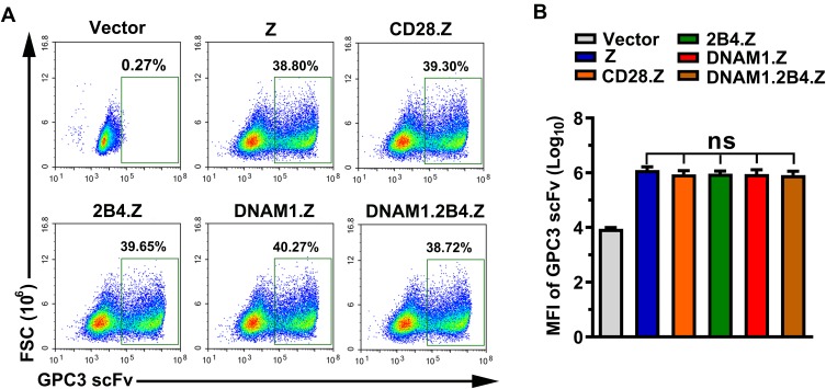
Figure 2.
Generation of CAR+ NK cells. (A) Representative images obtained using flow cytometry. After a 72-h infection with lentivirus, NK-92 cells were stained and analyzed by flow cytometry. The positive population represented CAR+ NK-92 cells. (B) Median fluorescence intensity (MFI) of GPC3-scFv on CAR+ NK-92 cells in each group (n=3; ns, no significance).

