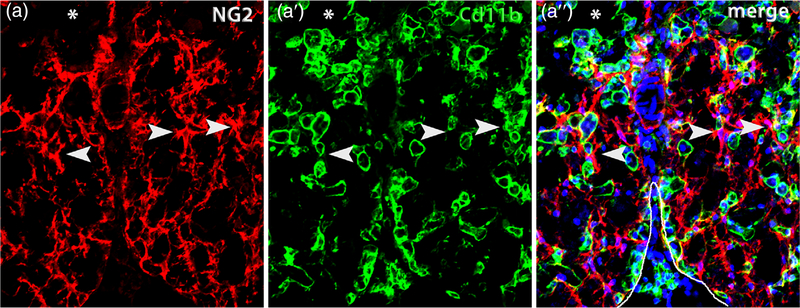
FIGURE 3.
OPCs closely intermingle with macrophages after spinal cord injury. Single channel and merged confocal images from ventral white matter bordering the lesion (*) immunolabeled for NG2 (red) (a) and Cd11b (green) (a’). The merged image (a”) is counterstained with Dapi (blue) and the ventral pial border is outlined in white. NG2+ OPCs and their processes are commonly adjacent to macrophages (examples indicated by arrowheads). This section is taken from the lesion epicenter at 14 days postinjury

