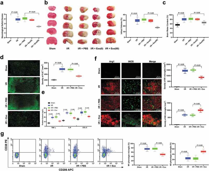
Figure 2.

hucMMSCs-exos ameliorate I/R induced M1 polarization and neuron deficiency. (a), neuron deficit scores (n = 20); (b), TTC staining (n = 5); (c), brain water content (n = 5) and (d) Fluo-Jade C staining (n = 5) were performed to determine mouse nerve system damage; E, ELISA was performed to determine pro-inflammation factors TNF-ɑ, IL-6, and CCL-2 protein contents; F, Immunofluorescence staining of M1 phenotype microglia marker iNOS and M2 phenotype microglia marker Arg 1 (n = 5); G, CD38+CD206− was categorized as M1 phenotype microglia while CD38−CD206+ was M2 microglia detected by flow cytometry (n = 5). Data are expressed as mean ± standard deviation and analyzed with one-way ANOVA, followed by Tukey’s multiple comparisons test. *p < 0.05. Three independent experiments were performed.
