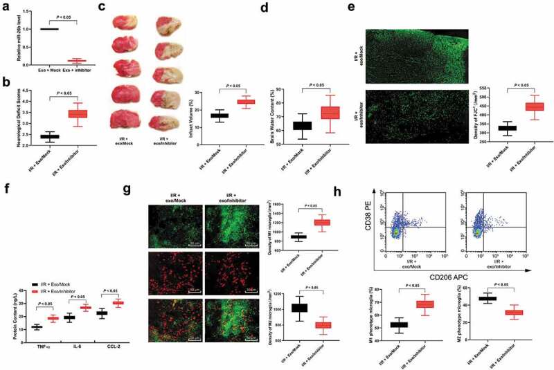
Figure 4.

hUCMSCs-exosomal miR-26b-5p downregulation reverses the effects of exosomes on microglia after I/R. (a), RT-qPCR was performed to determine miR-26b-5p expression in exosomes; (b), neuron deficit scores (n = 5), (c) TTC staining (n = 5), (d) brain water content (n = 5) and (e) Fluo-Jade C staining (n = 5) were performed to determine mice nerve system damage; F, ELISA was performed to determine pro-inflammation factors TNF-ɑ, IL-6, and CCL-2 protein contents; G, Immunofluorescence staining of M1 phenotype microglia marker iNOS, and M2 phenotype microglia marker Arg 1; H, CD38+CD206− was categorized as M1 phenotype microglia while CD38−CD206+ was M2 phenotype microglia detected by flow cytometry (n = 5). Data are expressed as mean ± standard deviation and analyzed with the unpaired t test (data in panels a, b, c, d, e and g) or one-way ANOVA (data in panels f and h), followed by Tukey’s multiple comparisons test. *p < 0.05. Three independent experiments were performed.
