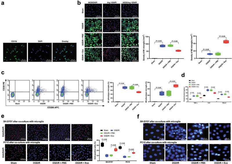
Figure 5.

Exosome treatment inhibits neuronal apoptosis induced by M1 microglia polarization following OGD/R. (a), CD11b immunofluorescent represents for microglia marker; (b), Immunofluorescence staining of M1 phenotype microglia marker iNOS and M2 phenotype microglia marker Arg 1; (c), CD38+CD206− was categorized as M1 phenotype microglia while CD38−CD206+ was M2 phenotype microglia detected by flow cytometry; (d), ELISA was performed to determine pro-inflammation factors TNF-ɑ, IL-6, and CCL-2 protein contents. Then, OGD/R and exosomes or PBS-treated microglia was co-cultured with SH-SY5Y or PC12 cells; EdU staining (e) and Hoechst 33258 staining (f) were performed to determine SH-SY5Y or PC12 cells viability and apoptosis induced by M1 microglia polarization in OGD/R. Data are expressed as mean ± standard deviation and data in panels A, B, C, D and E were analyzed with one-way ANOVA, followed by Tukey’s multiple comparisons test. *p < 0.05. Three independent experiments were performed.
