Figure 2.
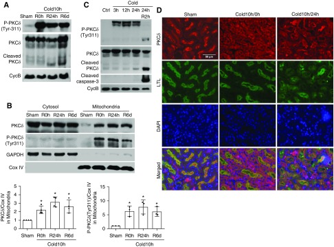
PKCδ is activated in cold-storage transplantation kidneys. The left kidney was collected from C57BL/6 donor mice for 10 hours of cold storage, followed by transplantation into syngeneic recipient mice for 0 hours, 24 hours, or 6 days. The right kidney of donor mice without cold storage–associated transplantation was used as sham control. (A) Representative immunoblots to show PKCδ cleavage and the phosphorylation of PKCδ at Tyr-311 in whole kidney lysate. Cyclophilin B (CycB) was used as the loading control. (B) Representative immunoblots and densitometry analysis of PKCδ and phosphorylated PKCδ in renal cytosolic and mitochondrial fractions. COX IV and glyceraldehyde-3-phosphate dehydrogenase (GAPDH) were used as loading controls of mitochondrial and cytosolic fractions, respectively. (C) Representative immunoblots show a rapid and sustained PKCδ (Tyr-311) phosphorylation during cold storage and proteolytic release of the PKCδ catalytic fragment by active caspase-3 during rewarming. Cyclophilin B (CycB) was used as the loading control. (D) Representative immunofluorescence images of PKCδ in renal outer medulla area showing PKCδ translocation. Paraffin-embedded sections were stained for PKCδ with Fluorescein labeled Lotus Tetragonolobus Lectin (LTL) as proximal tubular marker. Data in (B) are expressed as mean±SD (n=3–4). *P<0.05 versus sham control. Ctrl, control; DAPI, 4′,6-diamidino-2-phenylindole; R, reperfusion.

