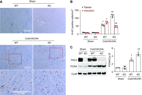
Figure 4.
PKCδ deficiency in donor kidneys increases tubular cell proliferation after transplantation. The left kidney was collected from PKCδ-KO or WT mice for 10 hours of cold storage, followed by transplantation into WT mice for 24 hours. The right kidney of donor mice without cold-storage transplantation was used as sham control. Kidneys were either fixed for histologic examination or lysed for immunoblot analysis. (A) Representative images of Ki67 immunohistochemistry. Bottom panels are enlarged images of the boxed areas in the middle panels. Scale bar, 0.2 mm. (B) Quantification of Ki67-positive tubular cells or interstitial cells. (C) Representative immunoblots and densitometry analysis of PCNA. Cyclophilin B (CycB) was used as loading control and used for normalization in densitometry. Data are expressed as mean±SD (n=4). *P<0.05 versus respective sham control group, #P<0.05 versus cold 10 hours/24 hours WT group.

