Figure 7.
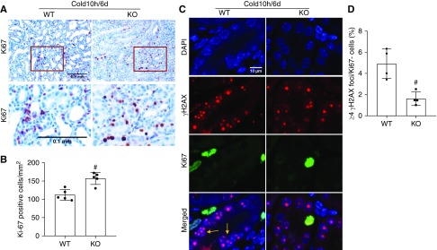
PKCδ deficiency improves tubular proliferation and decreases tubular senescence in life-supporting grafts. The left kidney was collected from PKCδ-KO or WT mice for 10 hours of cold storage, followed by transplantation into WT recipient mice. At day 5 post-transplantation, the native kidney of the recipient mouse was removed so the transplanted kidney became the life-supporting kidney. At day 6, the transplanted kidney was collected for analysis. (A) Representative images of Ki67 immunohistochemistry. Lower panels are enlarged images of the boxed areas in the upper panels. Scale bar, 0.1 mm. (B) Quantification of Ki67-positive tubular cells. (C) Representative double immunostainings of γH2AX (red) and Ki67 (green). Tubular cells with more than four γ-H2AX foci in nuclei and negative for Ki67 staining were considered senescent (arrows). (D) Quantification of cells with four or more γH2AX foci/Ki67− in kidney tubules. Data are expressed as mean±SD (n=4–5). #P<0.05 versus transplanted WT grafts. DAPI, 4′,6-diamidino-2-phenylindole.

