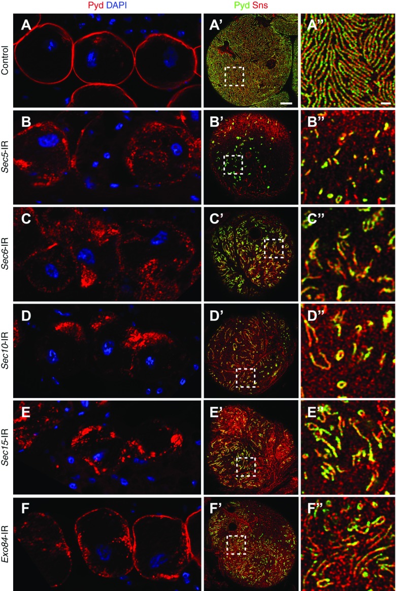
Figure 2.
Exocyst gene silencing disrupts localization of the NSD-associated proteins Pyd and Sns. (A) In control (wild-type) adult fly nephrocytes, Pyd (red) was tightly localized to the cell margin in the medial optical section, defining a highly regular, continuous circumferential ring. (A′ and A″) Immunofluorescence detection of Pyd (green) and Sns (red) in the plane of the cell surface showed a highly regular and uniform fingerprint-like pattern of distribution in wild-type control adult nephrocytes. Scale bar, 5 µm in A′; 1 µm in A″. (B–F, B′–F′, and B″–F″) Silencing of any of the exocyst genes resulted in a clear disruption of normal Pyd localization at the both cell margin and cell surface. Cell nuclei are labeled by 4′,6-diamidino-2-phenylindole (DAPI) staining (blue).

