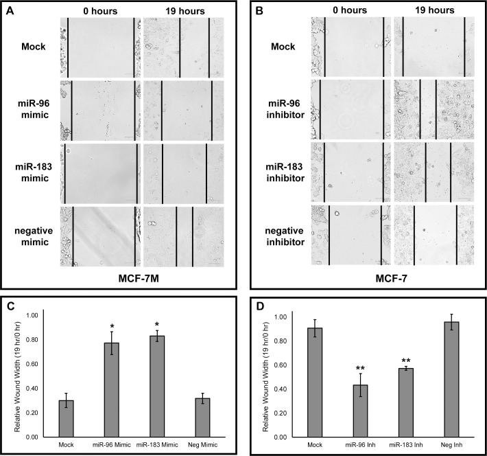
Fig 2. MicroRNAs miR-96 and miR-183 regulate cell migration.
(A) 50 nM of mirVana™ miRNA mimics for miR-96, miR-183, or Negative Control #1 were transfected into MCF-7M cells and (B) 50 nM of mirVana™ miRNA inhibitors for miR-96, miR-183, or Negative Control #1 were transfected into MCF-7 cells. Cells were allowed to grow for 24 hours post-transfection before each well was scraped in triplicate with a p-10 pipette tip. Images of wounds were taken using the EVOS FLoid Cell Imaging Station and are representative of three independent transfections. (C, D) Relative wound width was determined using ImageJ software (length of wound at 19 hours divided by length of wound at 0 hours) for MCF-7M cells treated with miRNA mimics and MCF-7 cells treated with miRNA inhibitors (Inh). Values expressed as averages ± SEM. *p < .05, **p < .01.

