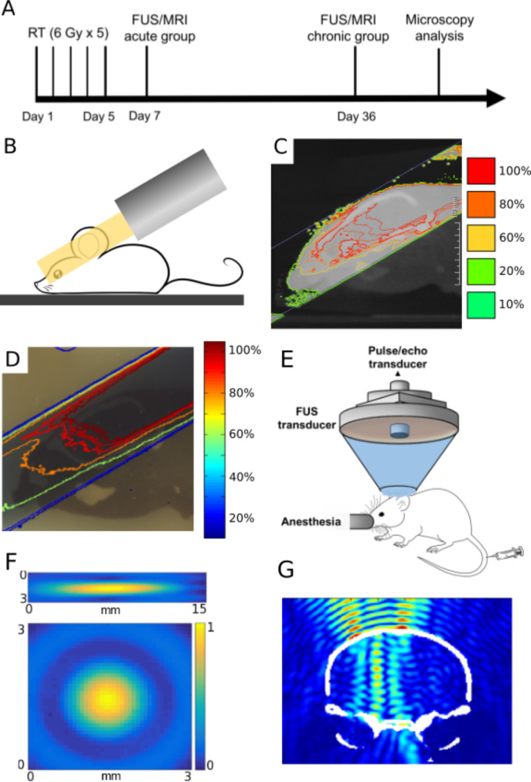
Figure 1.
Radiation and FUS experimental setup and timeline. (A) Experimental timeline of treatments and analyses. (B) Schematic of half brain radiation with a single beam using SARRP system. (C) Simulated dose distribution of radiation treatment plan in MuriPlan of the mouse brain. (D) Representative isodose lines measured with radiochromic film from irradiated mouse brain phantom. Both (C, D) are normalized to the maximum dose value in the brain. (E) Experimental setup of the FUS system used in all sonications. (F) The FUS focus was calibrated using an acoustic hydrophone to be 7.5 × 1 × 1 mm3. (G) Simulation of normalized FUS beam profile in the targeted half brain (coronal view). FUS,focused ultrasound; SARRP, Small Animal Radiation Research Platform.

