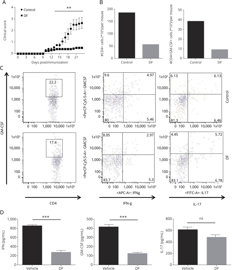
Figure 2. DF ameliorates experimental autoimmune encephalomyelitis and suppresses GM-CSF–producing Th1 cells in the CNS.
(A) Wild-type B6 mice were immunized with 200 μg of MOG35–55 and were treated with DF (treatment group) or placebo (control group) by oral administration starting on the day of immunization. Clinical signs were scored daily following a 0–5 scale. Data represent 1 of 3 experiments and the mean clinical scores ± SEM (n = 5 each group). (B) CNS-infiltrating cells from DF-treated and control mice were isolated on day 23 of disease. DF treatment significantly decreased total CD4+ T cells and GM-CSF–producing CD4+ T cells among CNS-infiltrating cells. (C) Cells were stained with GM-CSF, IFN-γ, and IL-17A antibodies and analyzed by flow cytometry. We found that GM-CSF was decreased in Th1 cells, but not Th17 cells, after DF treatment. (D) Splenocytes from both treated and control groups were collected and cultured/stimulated with 25 μg/mL MOG35–55 for 72 hours. Concentrations of GM-CSF, IFN-γ, and IL-17A in culture supernatants were measured by ELISA. There was significantly less GM-CSF and IFN-γ in treated mice compared with controls. **p < 0.01; and ***p < 0.001. One of 2 experiments is shown. DF = dimethyl fumarate; GM-CSF = granulocyte macrophage colony-stimulating factor; IL = interleukin; MOG = myelin oligodendrocyte glycoprotein; SEM = standard error of the mean.

