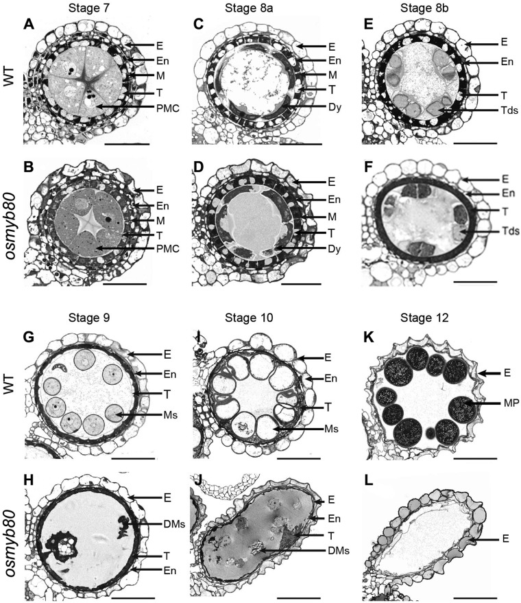
Fig. 3.
Histological features of anther development in the WT and osmyb80 mutant. WT (A, C, E, G, I, K) and osmyb80 (B, D, F, H, J, L) anthers at developmental stages 7–12 are shown. Scale bar = 20 μm. DMs, degenerated microspores; Dy, dyad cell; E, epidermis; En, endothecium; M, middle layer; MP, mature pollen; Ms, microspores; T, tapetal layer; Tds, tetrads.

