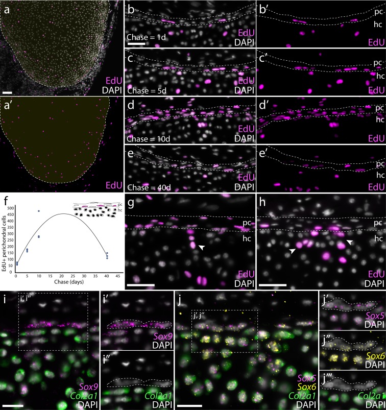
Figure 4. Proliferation of chondrocytes and recruitment of new chondrocytes from putative perichondral progenitors in skate hatchlings.
(a) Transverse sections through the metapterygium of skate hatchlings reveals a concentration of label-retaining (EdU+) cells around the periphery of the element, both in the cartilage and in the perichondrium, as well as sparse label-retaining cells in the center of the metapterygium. An EdU pulse-chase experiment reveals that the perichondrium is an expanding cell population. (b) 1 day chase reveals sporadic labelling of cells within the perichondrium, but an increase in the number of EdU+ perichondral cells after (c) 5- and (d) 10 day chases. (e) After a 40 day chase, reduction in the number EdU+ cells in the perichondrium points to label dilution over several rounds of cell division. (f) Sum total EdU+ perichondral cells from three adjacent 8 μm transverse paraffin sections through the metapterygium after EdU pulse and 1-, 5-, 10- and 40 day chase (n = 3 individuals per time point). (g) The occurrence of clusters of EdU+ chondrocytes immediately adjacent to EdU+ perichondral cells after 10 day and (h) 40 day chase points to the likely perichondral origin of these cells. mRNA in situ hybridisation by HCR for (i) Sox9 and Col2a1, and for (j), Sox5, Sox6 and Col2a1 reveals a population of perichondral cells that sit at the cartilage-perichondral boundary, and that co-express Sox9, Sox5 and Sox6 but not Col2a1. These cells (white dashed outline) are morphologically similar to the label-retaining perichondral cells identified in (b-e). In all images, plane of section as indicated in Figure 1i. hc, hyaline cartilage; pc, perichondrium. Scale bars: (a) 100 μm, (b-e), (g-h) 50 μm, (i-j) 25 μm.

