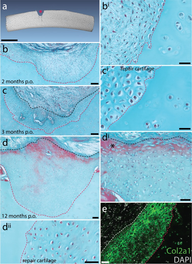
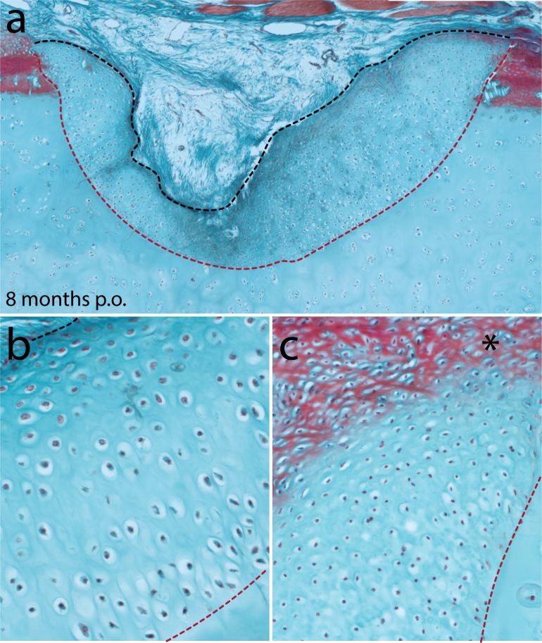
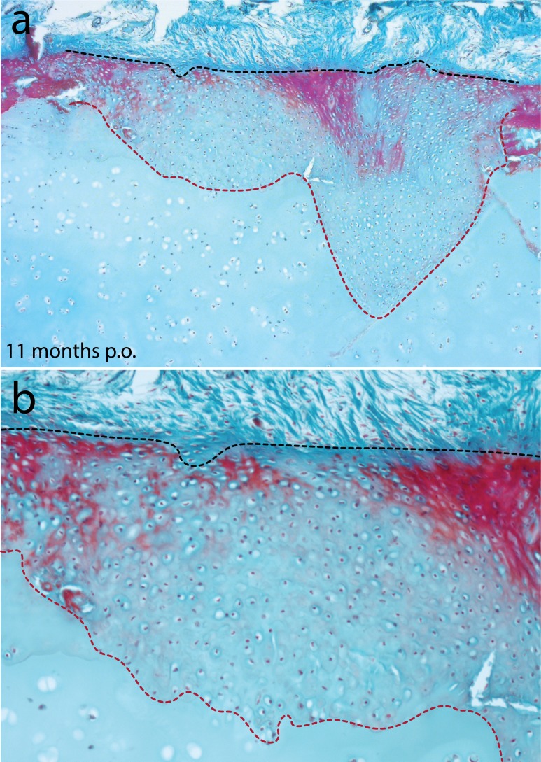
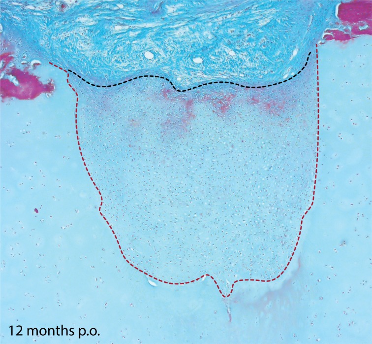
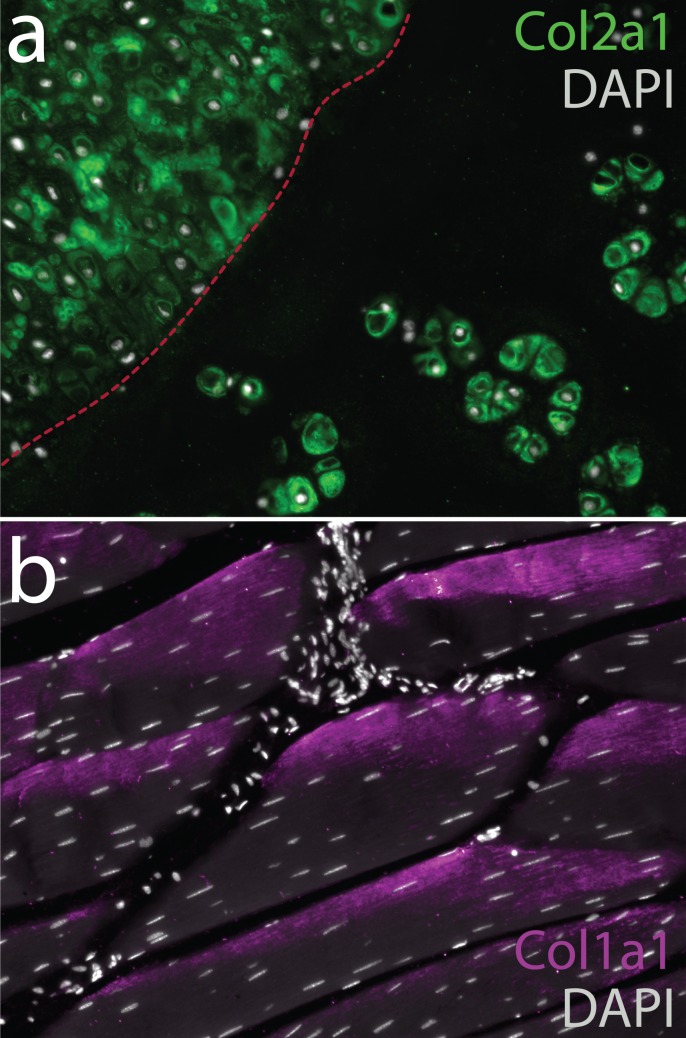
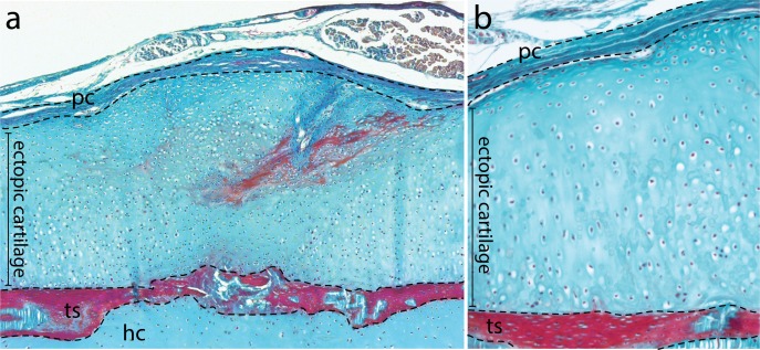
Figure 8. Spontaneous repair of hyaline cartilage in the skate.
(a) 3D reconstruction of a dissected metapterygium 2 weeks following experimental cartilage injury. Note the biopsy (red asterisk) has left a void of ~1/3 the diameter of the metapterygium. (b, bi) By 2 months post-operation (p.o.), the injury site has been filled with a fibrous connective tissue, and (c, ci) by 3 months p.o., this connective tissue begins to differentiate into cartilage. Note that the cells of the repair tissue adopt chondrocyte morphology, and the ECM of the repair tissue is integrated with adjacent cartilage. (d) By 12 months p.o., the injury site has been completely filled with repair cartilage. (di) Red staining of ECM at the surface of the repair tissue (*) could indicate the re-appearance of tissue with a perichondral-like nature, or the re-establishment of tesserae at the injured surface of the metapterygium. However, (dii) the vast majority of repair tissue is composed of typical hyaline cartilage. (e) Immunofluorescence reveals abundant type II collagen (Col2a1) in the ECM of repair cartilage. In (b-d), the red dashed line indicates the boundary of the biopsy, and the black dashed line indicates the extent of repair cartilage. In (e) the red dashed line indicates the boundary of the biopsy, and the white dashed line indicates the extent of repair cartilage. hc, hyaline cartilage; pc, perichondrium; ts, tesserae. Scale bars: (a) 1 cm, (b) 100 μm, (bi) 30 μm, (c) 100 μm, (ci) 30 μm, (d) 100 μm, (di) 50 μm, (dii) 50 μm, (e) 50 μm.

