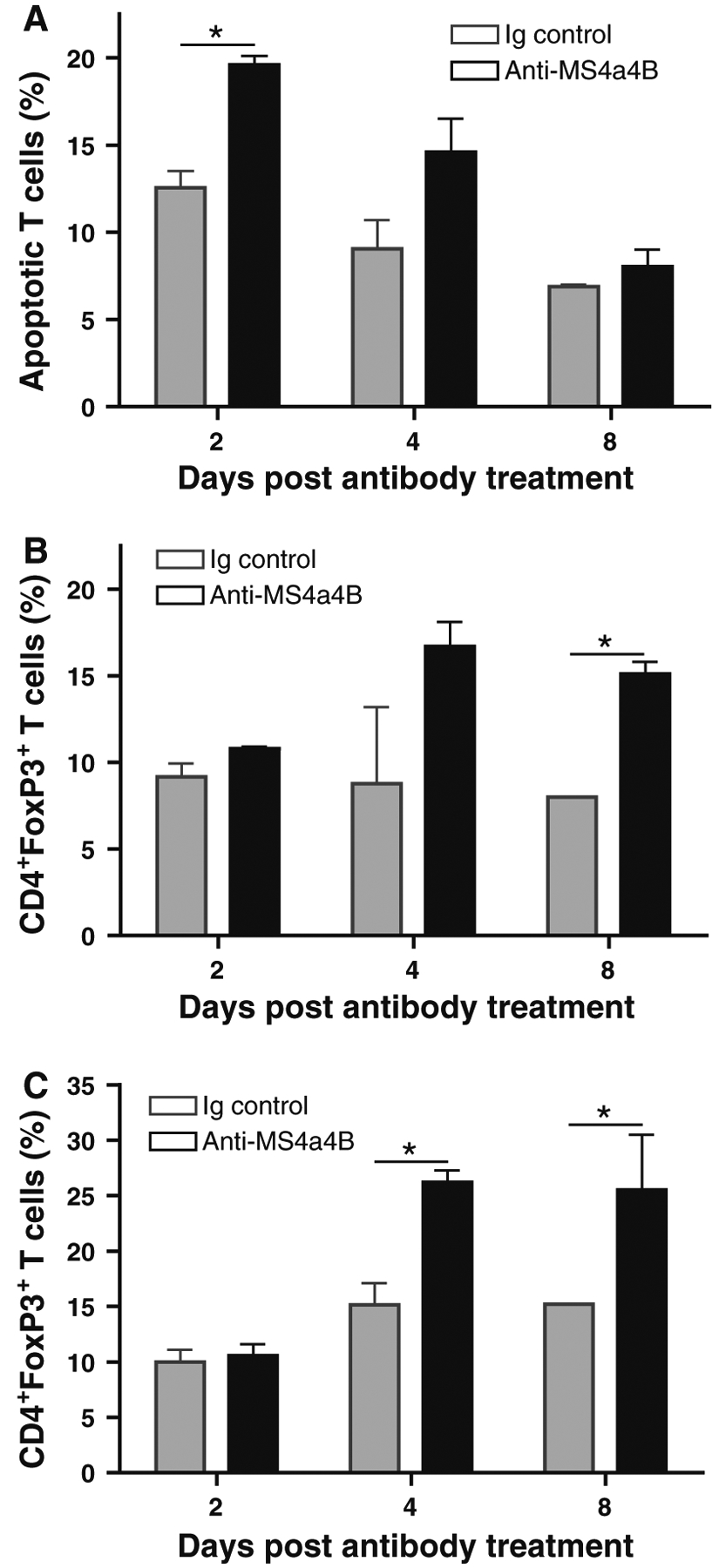
Fig. 6.

Anti-MS4a4B-mediated T cell apoptosis is accompanied by an elevated level of Treg cells. MOG-immunized C57BL/6 mice were injected with 1 mg/mouse anti-MS4a4B antibody or control rabbit IgG on day 9 post immunization. a Peripheral blood samples were collected from mice on days 2, 4 and 8 of antibody (or Ig) injection. After red blood cells were lysed, apoptotic T cells in samples were analyzed with Annexin V assay. Data are presented as mean ± SD of apoptotic T cell percentage (N = 3). b Treg cells in blood samples described in “a” were detected by flow cytometry with anti-CD4-FITC/anti-FoxP3-PE staining. Data are presented as mean ± SD of percentage of FoxP3+ T cells in CD4+ gate. c Treg cells in spleens from mice described in “a” were analyzed by flow cytometry as described in “b”. The results were presented as mean ± SD of percentage of FoxP3+ T cells in CD4+ gate (N = 3). A representative of two experiments is shown
