Fig. 3.

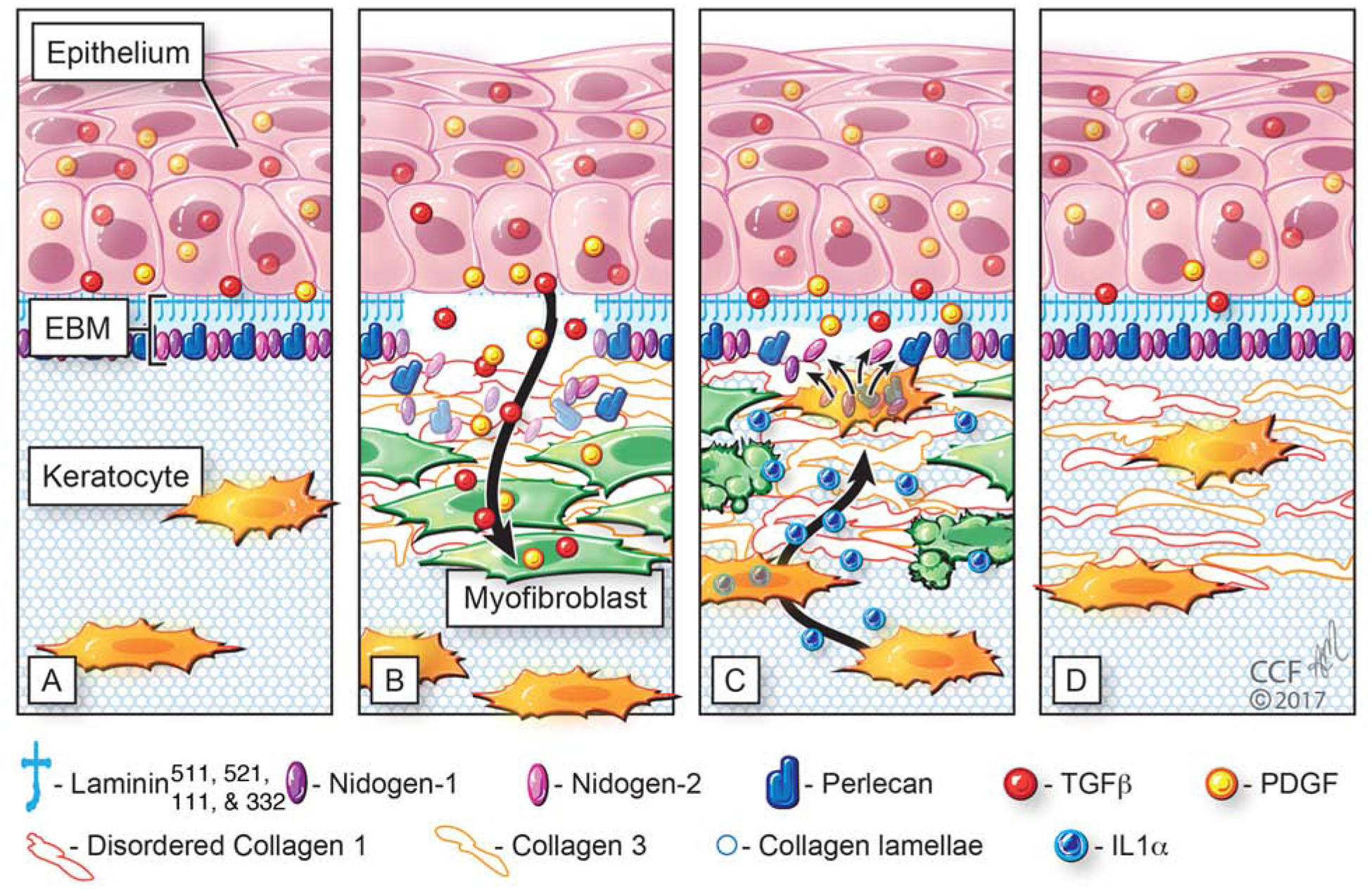
Transmission electron microscopy (TEM) images of rabbit corneas (e, epithelium; S, stroma). A. A clear cornea at two weeks after −4.5D PRK had normal regeneration of the EBM (arrows) with clear lamina densa and lamina lucida. B. A cornea with dense fibrotic haze at four weeks after −9.0D PRK showed a complete absence of regenerated EBM—although it is likely there is a nascent abnormal epithelial BM consisting of laminin and other components of EBM beneath the basal epithelial cells that doesn’t appear as the classic EBM on TEM. Also note the large numbers of myofibroblasts (cells indicated by arrowheads with prominent endoplasmic reticulum) in the anterior stroma surrounded by a disorganized extracellular matrix that would act as a barrier to keratocytes repopulating the anterior stroma.
