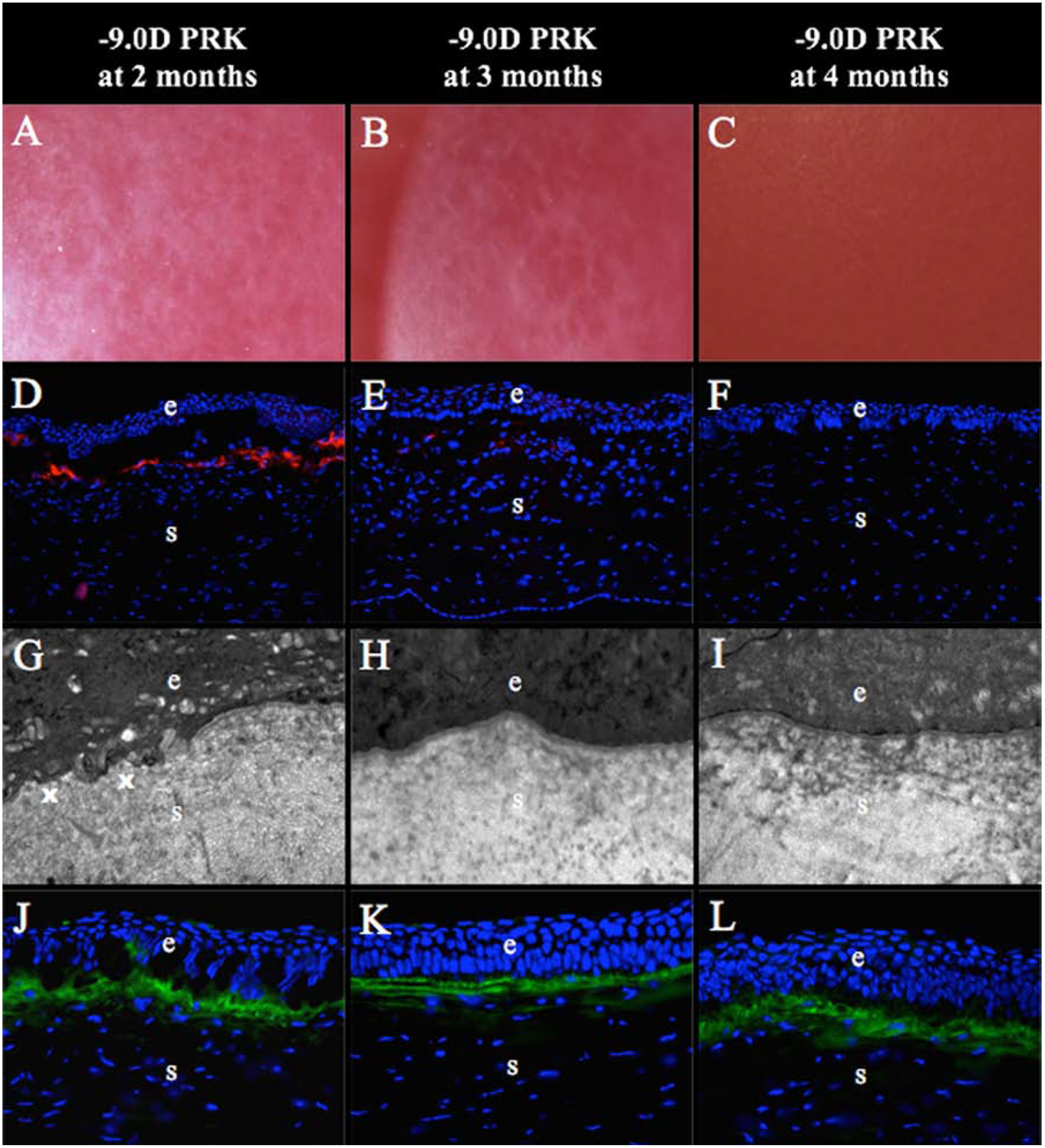
Fig. 5. Regeneration of the EBM at later time points after −9D PRK in rabbits.

(A) At two months after −9D PRK, all the corneas had developed clear areas called “lacunae” (arrows) within the confluent corneal fibrosis in the PRK ablation zone (mag 25X). (B) At three months after −9D PRK, lacunae (arrows) had enlarged and some had coalescedin all corneas (mag 25X). (C) At four months after −9D PRK, corneal transparency was fully restored in all corneas (mag 25X). (D) At two months after −9D PRK, large numbers of α-SMA+ myofibroblasts (arrowheads) were present in the fibrotic subepithelial stroma, although less than corneas at one month after PRK (see Fig. 4F) (mag 200X). (*) is artifactual detachment of the epithelium from stroma that occurred during cryostat tissue sectioning—likely due to defective EBM. (E) At three months after −9D PRK, there were only a few remaining α-SMA+ myofibroblast (arrowheads) in the superficial stroma within the PRK ablated zone (mag 200X). (F) At four months after −9D PRK, there were no remaining α-SMA+ myofibroblasts in the superficial stroma (mag 200X). (G) At two months after −9D PRK, transmission electron microscopy (TEM) of the excimer laser ablated zone showed areas of fully-regenerated EBM with lamina lucida and lamina densa (arrows) adjacent to areas without normal EBM (x) (mag 23,000X). (H) At three months after −9D PRK, normal EBM (arrows) was regenerated in the PRK ablated zone of all corneas (mag 23,000X). (I) At four months after −9D PRK, normal EBM was present in the PRK ablated zone in all corneas (mag 23,000X). (J) At 2 months after −9D PRK, large amounts of collagen type III (COL3, arrows) were detected in superficial stroma of the PRK ablated zone (mag 400X). The level of COL3 deposited in the subepithelial stroma appeared unchanged at three months (K) and four months (L) after −9D PRK (mag 400X). Blue is DAPI staining of cell nuclei; (e) epithelium; (s) stroma. Reprinted with permission from Marino GK, Santhiago MR, Santhanam A, Lassance L, Thangavadivel S, Medeiros CS, Torricelli AAM, Wilson SE. Regeneration of defective epithelial basement membrane and restoration of corneal transparency. J. Ref Surg. 2017;33:337–346.
