
Figure 1. MAG and myelin block activation of Rap1 by BDNF.
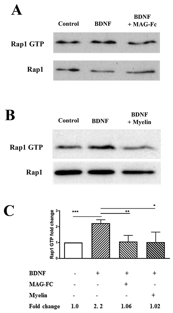
(A, B) Western blots of P5-6 CGN treated with BDNF (200 ng/ml) for 20 minutes, or with MAG-Fc (20 μg/ml) or CNS myelin (20 μg/ml) for 20 minutes prior to the addition of BDNF (n=4 for experiments with MAG-Fc, n=3 for experiments with myelin). Lysates were used for Rap1 activation assays detecting GTP-bound Rap1, and total Rap1 was assessed using input samples. (C) Quantification of Rap1 activation, where Rap1 GTP levels were normalized to the total Rap1 levels in the input lysate. Graphs depict average fold changes ± SEM (***p < 0.001, **p < 0.01, *p < 0.05 one-way ANOVA with Bonferroni’s multiple comparisons test).