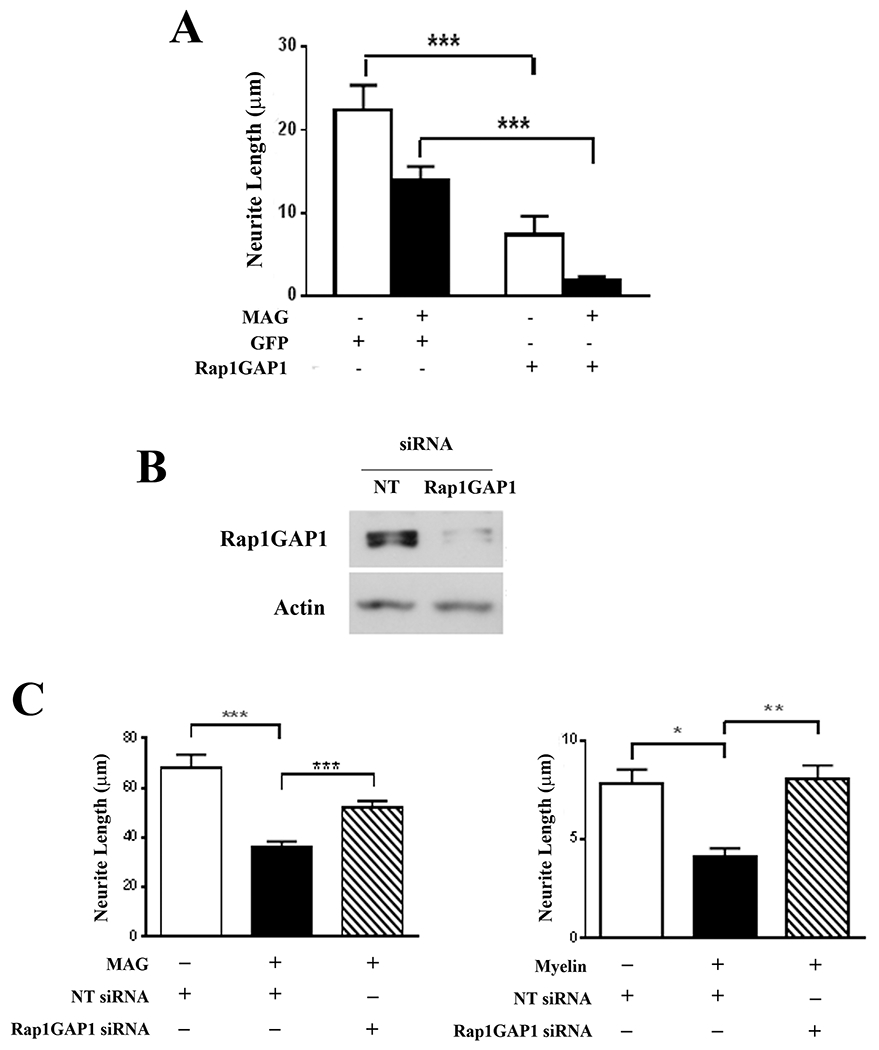
Figure 5. Overexpression of Rap1GAP1 inhibits neurite outgrowth while Rap1GAP1 knockdown blocks MAG-mediated inhibition of neurite outgrowth.

(A) Quantification of neurite outgrowth for P1-3 hippocampal neurons that were infected with adenoviruses expressing GFP, or GFP and Rap1GAPI. Neurons were incubated for 24 hours and transferred to control or MAG-expressing CHO cell monolayers. Graph depicts average length of the longest neurite per neuron ±SEM for approximately 50 neurons per treatment that were positive for both GFP and βIII tubulin (3 independent experiments, ***p < 0.001, one-way ANOVA with Bonferroni’s multiple comparisons test). (B) Western blots of P1-3 cortical neurons that were transfected with either non-targeting (NT) or Rap1GAP1 siRNA and incubated for 24 hours. (C) Quantification of neurite outgrowth for P1-3 cortical neurons that were transfected with either non-targeting (NT) or Rap1GAP1 siRNA and transferred to CHO cell monolayers or CNS myelin substrates. Graphs depict average length of the longest neurite per neuron ±SEM for approximately 100 neurons per treatment (3 independent experiments, *p < 0.05, **p < 0.01, ***p < 0.001, one-way ANOVA with Bonferroni’s multiple comparisons test).
