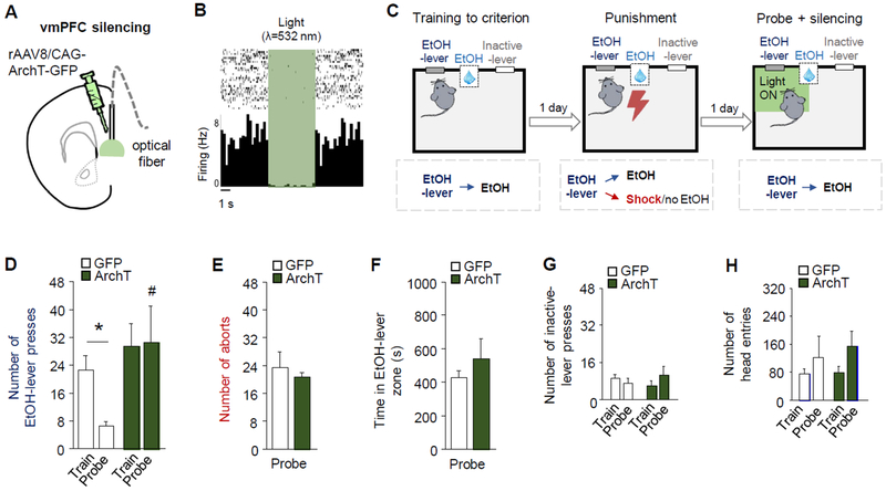
Figure 3: Closed-loop vmPFC-silencing reverses punished-suppression.
(A) Optical fibers were bilaterally directed at vmPFC neurons transfected with rAAV8/CAG-ArchT-GFP (ArchT). (B) In vivo recordings showing vmPFC neuronal inhibition in response to green light. (C) Green light was shone to silence vmPFC neurons when mice approached the EtOH-lever during probe testing. (D) Difference in EtOH-SA between ArchT and GFP groups (two-way ANOVA, group main effect: F(1,12)=5.91, P=. 03). Significant suppression of EtOH-lever pressing during the probe test in GFP controls (mean=22.6 train, 9.3 punishment, 6.6 probe) (paired t-test: t(6)=4.09, P<0.01), but not vmPFC-silenced ArchT group (mean=29.5 train, 7.4 punishment, 30.6 probe) (paired t-test: t(6)=0.08, P=0.93), such that the rate on the probe test was significantly higher in the ArchT group than the controls (unpaired t-test: t(12)=2.30, P=0.04). GFP and ArchT groups did not differ in (E) aborts (unpaired t-test: t(11)=0.52, P=0.61) or (F) cumulative duration in the light-on zone (unpaired t-test: t(12)=0.88, P=0.40). (G) The rate of inactive-lever pressing was unaffected by punishment (two-way ANOVA, session main effect: F(1,12)=.001, P=.98), but the ArchT group pressed the inactive-lever more than GFP (group main effect: F(1,12)=7.93, P=.02). (H) The rate of head-entries into the reward-receptacle was unaffected by punishment (two-way ANOVA, session main effect: F(1,12)=2.75, P=.12) or group (group main effect: F(1,12)=2.75, P=.12). Data are means ± SEM from n=7 mice/group. *P<.05 versus train in the ArchT group; #P<.05 versus probe in GFP controls.

