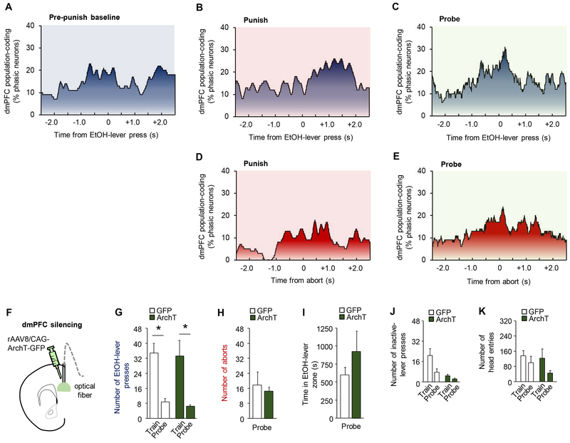
Figure 4. dmPFC encoding of events related to punished-suppression.
dmPFC neurons did not display clear representations of EtOH-lever presses (A) during the baseline period before punishment, nor coding of presses (B) or aborts (C) in the punishment period itself, though there was evidence of some level of press-related coding during the probe trial (D-E). (F) Optical fibers were bilaterally directed at dmPFC neurons transfected with rAAV8/CAG-ArchT-GFP (ArchT). Green light was shone to silence vmPFC neurons when mice approached the EtOH-lever during probe testing. (G) Significant suppression of EtOH-lever pressing during the probe test (two-way ANOVA, session main effect: F(1,12)=25.50, P=.0003) that did not differ between GFP and ArchT groups (GFP mean= 33.7 train, 8.2 punishment, 9.4 probe; ArchT mean=33.3 train, 6.6 punishment, 6.7 probe) (group main effect: F(1,12)=.053, P=.82). (H) GFP and ArchT groups did not differ in the rate of aborts (unpaired t-test: t(12)=0.43, P=0.67) or (I) time in the light-on zone (unpaired t-test: t(12)=1.09, P=0.30). (J) The rate of inactive-lever presses was higher in GFP versus ArchT groups (two-way ANOVA, group main effect: F(1,12)=14.37, P=.003) and decreased in all animals following punishment (session main effect: F(1,12)=5.10, P=.04) (K) Rate of head-entries decreased for both groups following punishment (two-way ANOVA, session main effect: F(1,12)=14.21, P=.049), but did not differ between groups (group main effect: F(1,12)=.78, P=.39). Data are means ± SEM from a total of n=129 (punished session) and 124 (probe tests) neurons in n=20 mice for A-E, and n=7 mice/group for F-K. *P<.05 versus train in the ArchT group; #P<.05 versus probe in GFP controls.

