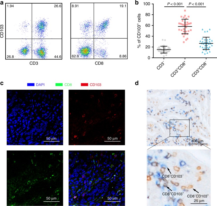
Fig. 1. Identification of CD103+CD8+ T cells in human gastric cancer tissues.
a Representative images from flow cytometry showing the co-expression of CD103 with the T cell surface markers CD3 and CD8 (gated on the CD45+ subset). b Scatter plot showing the fractions of CD103+ immune cells in gastric cancer (n = 36). c Representative immunofluorescence staining of CD8 and CD103 in gastric cancer tissues. d Representative dual immunohistochemical staining of CD8 and CD103 in gastric cancer tissues. Bar plots show the mean ± SD.

