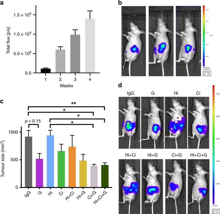
Fig. 1. Primary tumour volume.
a Bioluminescence imaging of mice over the first 4 weeks after cell implantation demonstrated that tumours grew at similar rates over that period, and importantly, that the tumour sizes were similar in all mice at the initiation of treatment. b Representative photographs of bioluminescent imaging at 4 weeks, showing that cancer cells have metastasised (luminescent signal separate from primary tumour). c Effects of HGF/c-MET inhibition ± gemcitabine on tumour volume. The combination of c-Met inhibitor + gemcitabine (Ci + G) and HGF-neutralising antibody + c-MET inhibitor + gemcitabine (triple therapy, Hi + Ci + G) significantly reduced tumour size. **p < 0.01 Hi + Ci + G vs. IgG; *p < 0.05 Hi + Ci + G vs. Hi, or Ci + G vs. IgG, or Ci + G vs. Hi; n = 6 mice per group. d Representative photographs of bioluminescent imaging at the end of the 6-week treatment period, showing that triple therapy (Hi + Ci + G) and Ci + G significantly inhibited tumour growth, as determined by both signal intensity and area. Spectrum ranges from weak (blue) to strong (red).

