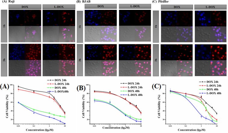
Figure 2.
Endocytosis of DOX and cell viability. Upper panels show confocal microscopy results of DOX signal from (A) Raji cell (B) BJAB cell and (C) Pfeiffer cell after incubated by DOX or L-DOX for 1 h or 3 h. (40×). The lower panels show the cell viability of (A) Raji cell, (B) BJAB cell, and (C) Pfeiffer cell in vitro with DOX and L-DOX. CCK-8 assays were performed. Data shown are means ± SD from three separate experiments (n = 3).

