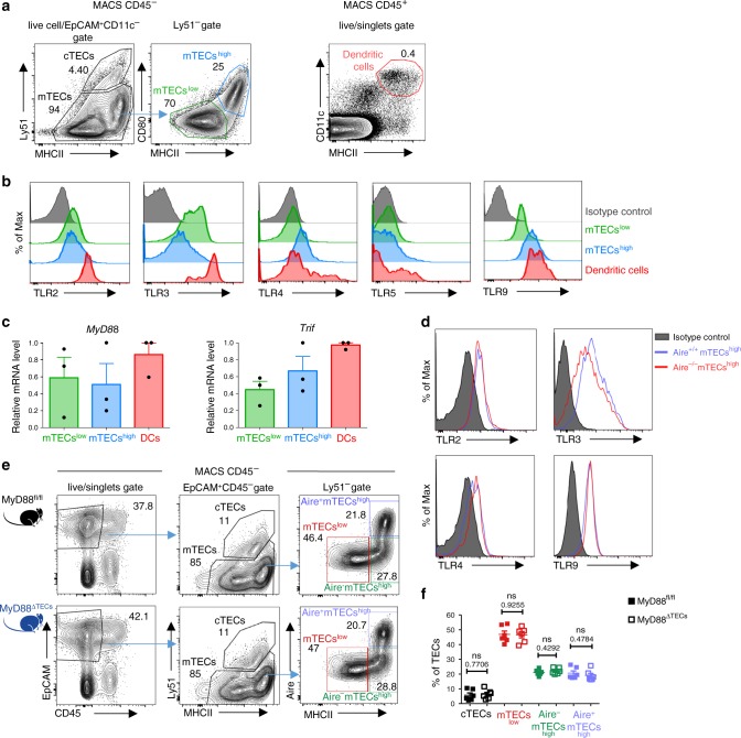
Fig. 1. mTECs express a set of TLRs and their signaling adaptors independently of Aire.
a Gating strategy used for the analysis of TEC populations and general thymic conventional DCs. MACS enriched CD45– and EpCAM+CD11c– pre-gated cells were further divided into cTECs (Ly51+), mTECslow (MHCIIlowCD80low), and mTECshigh (MHCIIhighCD80high). Thymic conventional DCs were gated as CD11c+MHCII+ from the CD45+ fraction. A more detailed gating strategy is found in Supplementary Fig. 1a, b. b Representative flow cytometry histograms of TLR expression on mTECs and DCs isolated from the thymus (n = 3 independent experiments). c MyD88 and Trif mRNA expression is determined by qRT-PCR from FACS sorted mTECs and DCs. The expression is calculated relative to Casc3 and normalized to the highest value within each experiment=1 (mean ± SEM, n = 3 samples). d Representative flow cytometry histograms of TLR expression on mTECs from Aire+/+ and Aire–/– mice, (n = 3 independent experiments). e Representative comparative flow cytometry plots of different TEC subpopulations in MyD88fl/fl and MyD88ΔTECs mice. f Quantification of TEC frequencies from plots in e (mean ± SEM, n = 6 mice). Statistical analysis was performed by unpaired, two-tailed Student’s t-test, p-values are shown. ns = not significant.

