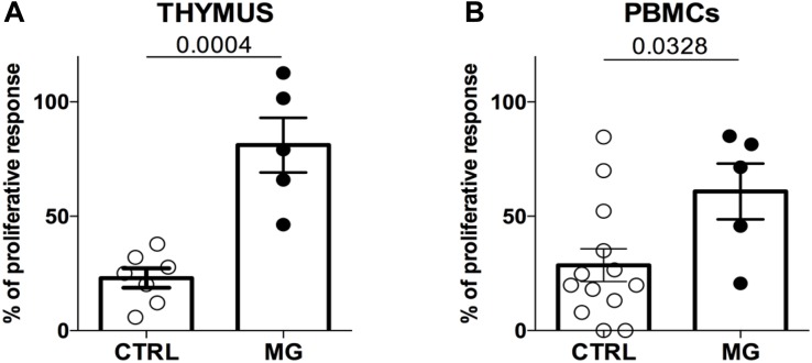
FIGURE 1.
The suppression function is more impaired in the thymus than in the periphery in MG patients. Percentages of proliferation of Tconv in co-culture with Treg (ratio 1:1) from control individuals (CTRL) or patients with myasthenia gravis (MG) using cells derived from the thymus (A) or from PBMCs (B). Data represent the mean ± standard error of the mean. Statistical test: Two-tailed t-test.

