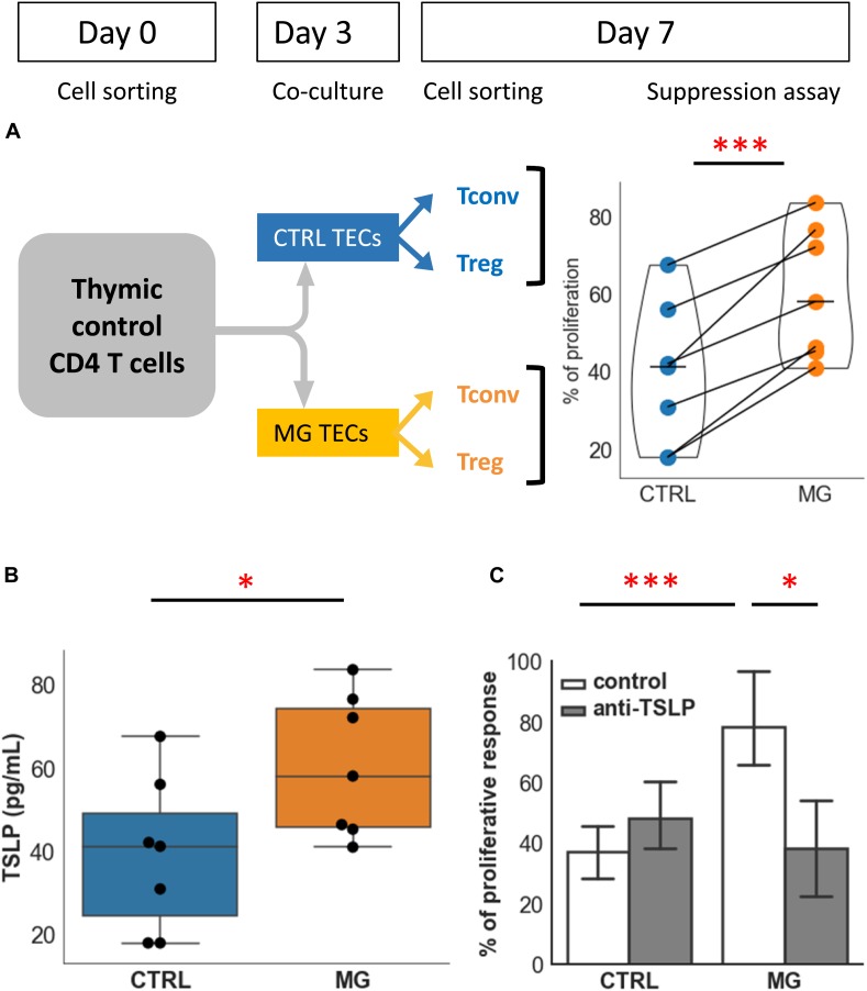
FIGURE 4.
TECs contribute to the functional defect of Treg: role of TSLP. (A) Percentages of proliferation of T conv in co-culture with Treg following co-culture of CD4 + cells from control individuals with TECs either from control individuals (CTRL, blue) or from MG patients (MG, orange). The experimental protocol is explained in the top left panel. (B) Production of TSLP measured in supernatants of cultured TEC from control individuals (CTRL, blue) or MG patients (MG, orange). (C) Percentage of proliferation of Tconv in co-culture with Treg following co-culture with TEC from control individuals (CTRL) or MG patients as described in (A), in the absence or presence of a TSLP neutralizing antibody. Statistical tests: two tailed paired t-test in (A,C), two-tailed Mann-Whitney in (B) (∗p ≤ 0.05; ∗∗∗p ≤ 0.005).

