Refer to full videos in the
Supplementary Material. Figure images are video stills.
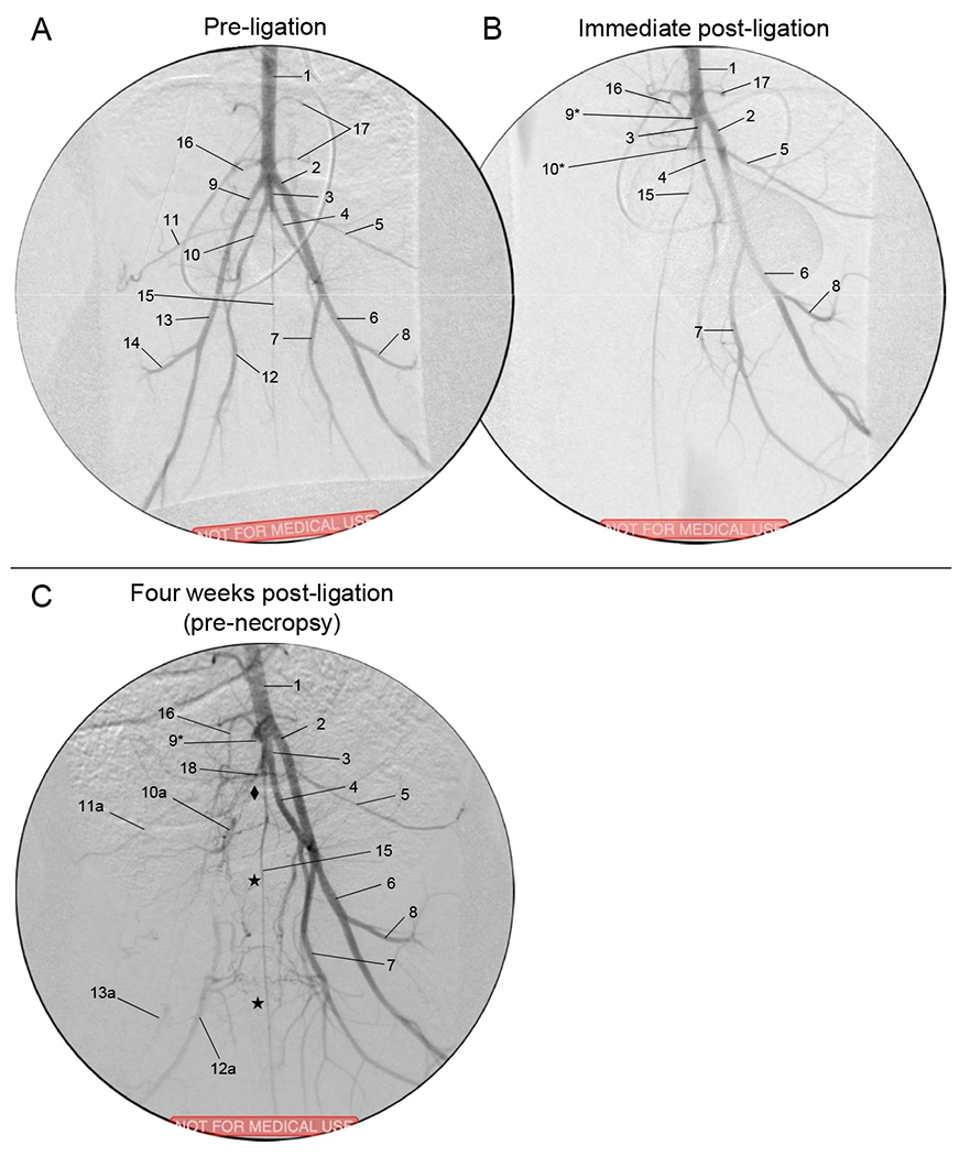
(A) Pre-ligation (baseline).
(B) Immediate post-ligation angiogram. The REIA, RIIA, and all distal vessels are absent.
(C) Thirty days later, pre-necropsy. Stars (★) indicate cross-over collaterals from LPFA which reconstitute the RPFA and then the RSFA; diamond (♦□) indicates collaterals from CIIT/LIIA which reconstitute the RIIA.
Aorta
Left external iliac artery
Common internal iliac artery trunk
Left internal iliac artery
Left lateral circumflex iliac artery
Left superficial femoral artery
Left profunda femoris
Left lateral circumflex femoral artery
Right external iliac artery (*ligated stump)
Right internal iliac artery (*ligated stump)
Right lateral circumflex iliac artery
Right superficial femoral artery
Right lateral circumflex femoral artery
Middle sacral artery
Inferior mesenteric artery
Lumbar branch
Posterior branch of CIIT

