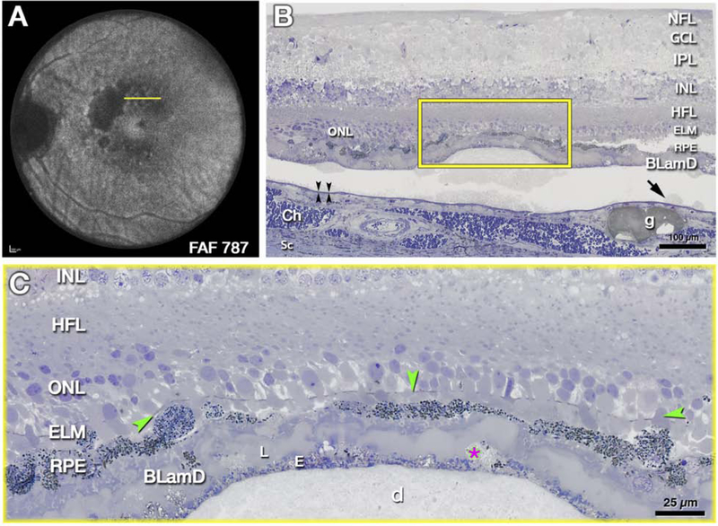
Figure 4: External limiting membrane (ELM) approaches druse apex due to photoreceptor shortening.
The histology in Fig 4 supports clinical imaging in Fig 2, 3 and illustrates changes in photoreceptor layers above a large druse as the atrophic process begins. A. Ex vivo imaging of the left eye of 85-year-old white female donor with geographic atrophy. Areas of absent autofluorescence signal (λ=787 nm) indicate complete retinal pigment epithelium (RPE) and outer retinal atrophy (cRORA). Yellow line crosses an area of mottled autofluorescence shown by histology. B, C. Submicrometer epoxy sections of osmium tannic acid paraphenylenediamine post-fixed tissue, stained with toluidine blue, at the plane indicated in A. B. Retina, RPE, and basal laminar deposit (BLamD) are artifactually detached from Bruch’s membrane (black arrowheads) at a soft druse and surrounding basal linear deposit (arrow). Framed area is magnified in C. NFL, nerve fiber layer; GCL, ganglion cell layer; IPL, inner plexiform layer; HFL, Henle fiber layer; ONL, outer nuclear layer; ELM, external limiting membrane; Ch, choroid; Sc, Sclera. g, Friedman lipid globule. C. An intact ELM (green arrowheads) skims close to the druse (d) apex, because photoreceptor outer segments are absent and inner segments are markedly shortened. RPE atop the druse is dysmorphic or absent. BLamD is thick with sublayers (L, late, E, early), including basal mounds (asterisk). Histology and figure prepared by J.D. Messinger DC from the Project MACULA resource http://projectmacula.cis.uab.edu/

