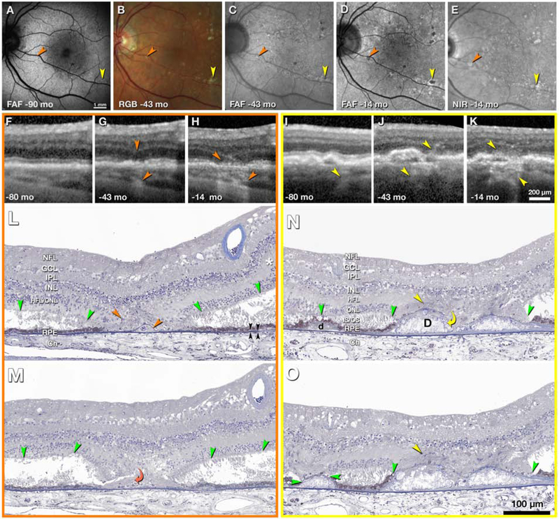
Figure 5. Evolution and clinicopathologic correlation of druse-associated atrophy.
Time points of clinical images are shown as months before death of this 90-year-old white woman. A-E, en face imaging shows developing atrophic spots (orange and yellow arrowheads). FAF, fundus autofluorescence (excitation wavelengths 488, 535–585, 532 nm in A,C,D respectively); RGB, color photograph; NIR, near-infrared reflectance. F-H, I-K, optical coherence tomography (OCT) B-scans through the orange and yellow atrophic spots, respectively. Upper and lower arrowheads in G-H and I-J indicate hyperreflectivity corresponding to gliosis and hypertransmission into the choroid, respectively; L-N, histologic sections through yellow and orange atrophic spots. In atrophic areas retina is attached to posterior tissues. Outside atrophic areas there is artifactual bacillary layer detachment. The HFL, in areas of minimal photoreceptor degeneration (asterisk in L), is pale-stained and ordered. In areas of photoreceptor loss, the HFL is stained medium-gray, with disordered fibers and evidence of Müller cell bodies (orange and yellow arrowheads), signifying gliosis. Green arrowheads, external limiting membrane (ELM). Black arrowheads, Bruch’s membrane. L, M, histologic sections 60 μm apart through the orange atrophic spot. In L is a base-down triangle of gliosis (upper arrowhead) and an area of absent ONL bounded by two ELM descents. The prior presence of a druse is indicated by calcific nodules under a blue-stained line of persistent BLamD (lower arrowhead in L). In M, the curved arrowhead indicates where processes from the HFL enter under the BLamD. N, O, two histologic sections 30 μm apart through the yellow atrophic spot. In the center are two drusen (D) with absent RPE and containing large calcific nodules. Through an interruption in the BLamD, gliotic processes enter from the HFL (yellow curved arrow, N). In the HFL are presumed Müller cell bodies (yellow arrowheads, N, O). On the left side of N is a small druse (d) with continuous RPE and ELM. In O, the ELM has descended onto the druse apex, which is covered with persistent BLamD. The RPE is absent. BLamD, basal laminar deposit. NFL, nerve fiber layer; GCL, Ganglion cell layer; IPL, inner plexiform layer; INL, inner nuclear layer; OPL, outer plexiform layer; HFL, Henle fiber layer; ONL, outer nuclear layer; IS/OS, inner and outer segments; RPE, retinal pigment epithelium; Ch, choroid. Prepared by J.D. Messinger DC and L. Chen MD PhD.

