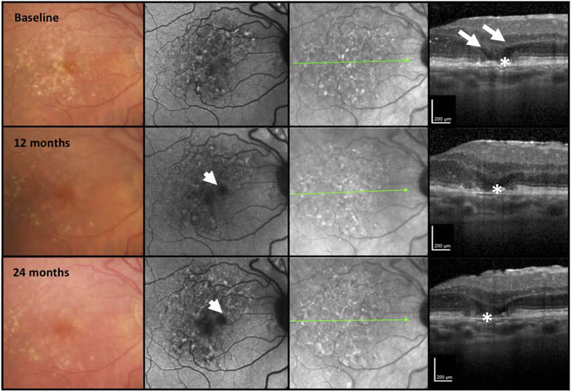
Figure 7. Multimodal imaging of an AMD case illustrating retinal pigment epithelium loss without choroidal hypertransmission over 24 months.
First column = color fundus photograph (CFP), second column = fundus autofluorescence (FAF), third column = near infrared reflectance (NIR), fourth column = optical coherence tomography (OCT) B-scan.
The right macula of a case demonstrating large drusen and pigmentary abnormalities on the CFP. At baseline the FAF demonstrates a mottled hypo- and hyperautofluorescent signal. The NIR also presents a mottled reflectance. The OCT demonstrates minimal subsidence of the inner nuclear layer (INL), outer plexiform layer; (OPL) but obvious hyporeflective wedges in Henle’s fiber layer (HFL) (large arrows). There is RPE attenuation (*) without obvious hypertransmission into the choroid. At 12 and 24 months, whilst there is increasing hypoautofluorescence on FAF imaging and progressive loss of the RPE (*) and a bare Bruch’s membreane (BrM). There is minimal hypertransmission into the choroid on the OCT, where greater hypertransmission would be expected to accompany the RPE loss. At no time point in this illustration were all the criteria met for iRORA.

