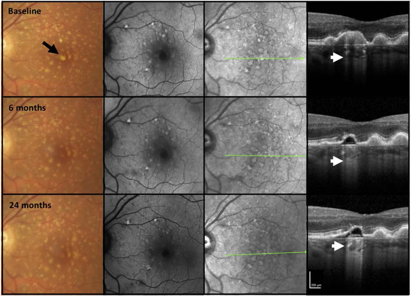
Figure 9. Multimodal imaging of an AMD case illustrating marked choroidal hypertransmission despite relatively intact retinal pigment epithelium and persistence of the druse contour.
First column = color fundus photograph (CFP), second column = fundus autofluorescence (FAF), third column = near infrared reflectance (NIR), fourth column = optical coherence tomography (OCT) B-scan
The left macula of a case illustrating large drusen on CFP with some hypopigmentation at baseline (black arrow). FAF and NIR do not demonstrate areas of hypoautofluorscence on FAF or reduced NIR, respectively. On OCT at baseline a druse displays hypertransmission into the choroid (bar code appearance, white arrow). There is no subsidence of the the inner nuclear layer (INL), outer plexiform layer; (OPL), and the RPE is preserved, thus not fulfilling all criteria for iRORA. At 6 months, the druse has reduced in size and appears hyporeflective but the overlying RPE remains intact above it, yet there is hypertransmission of the signal into the choroid (small arrow). All the criteria for iRORA are still not met. Over time the druse contour and marked hypertransmission persist (small arrow), and internal reflectivity partially fills the druse, the RPE is now becoming attenuated, although still with potentially residual basal laminar deposit (BLamD). At 24 months, all the criteria for iRORA have been met. Despite the hypertransmission there is no obvious hypoautofluorescence on FAF, although this area is masked by luteal pigment. No hyporeflective areas are seen on NIR, nor is there any corresponding GA on CFP.

