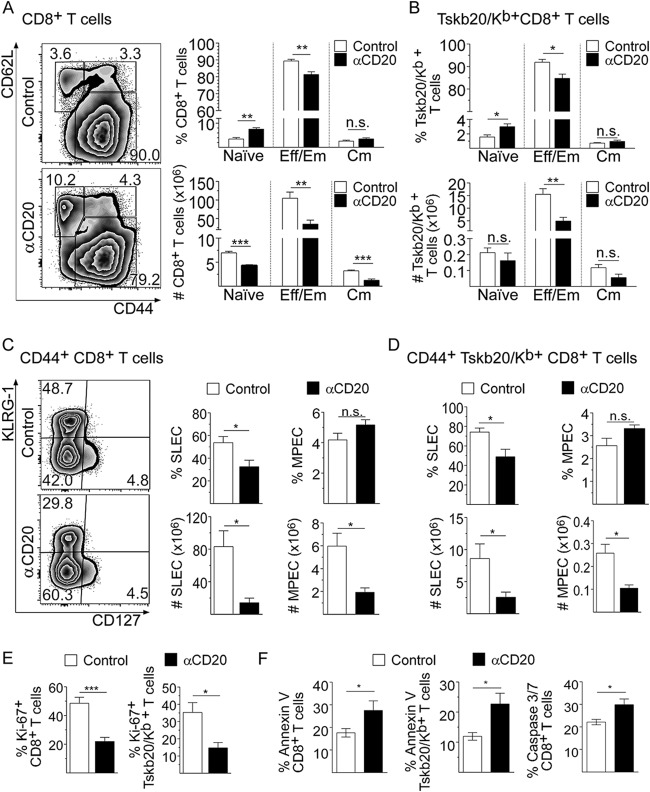
FIG 2.
Anti-CD20 treatment decreased effector and memory CD8+ T cell number and compromised their survival and proliferation. Mice injected with isotype control (control; in white bars) or anti-CD20 (αCD20; in black bars) mAb were infected with 5,000 trypomastigotes of T. cruzi strain Tulahuén. Splenic cells were obtained at 20 dpi and analyzed by flow cytometry. (A) Representative plots of CD62L versus CD44 expression on CD8+ T cells. (A and B) Statistical analysis of the frequency and number of naive (CD62Lhi CD44lo), effector memory/effector (Eff/Em) (CD62Llo CD44hi) and central memory (Cm) (CD62Lhi CD44hi) cells of (A) total CD8+ and (B) Tskb20/Kb+ CD8+ T cells. (C) Representative plots of KLRG-1 versus CD127 expression on CD44+ CD8+ T cells. (C and D) Statistical analysis of the frequency and number of short-lived effector cells (SLEC: KLRGhi CD127lo) and memory precursor effector cells (MPEC: KLRG1lo CD127hi) of (C) total CD44+ CD8+ and (D) CD44+ Tskb20/Kb+ CD8+ T cells. (E) Bar graphs representing the frequency of Ki-67+ cells on gated total CD8+ or Tskb20/Kb+ CD8+ T cells. (F) Bar graphs representing the frequency of apoptotic annexin V+ 7ADDneg cells in gated CD8+ or Tskb20/Kb+ CD8+ T cells and active caspase 3/7+ Sytoxneg cells in total CD8+ T cells. Results are representative of three (A to D) and two (E and F) independent experiments with 4 to 5 mice per group each. All P values were calculated with the two-tailed t test.

