FIG 1.
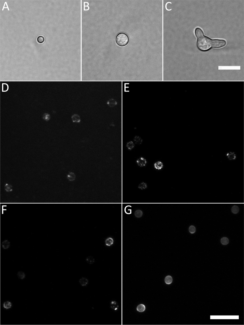
Cell morphotypes and labels used in this study. (A to C) The three developmental stages (morphotypes) analyzed in this study: dormant conidium (A), isotropically grown conidium (B), and germling (C) shown by bright-field microscopy. (D to G) Labeling of dormant conidia with fluorescently labeled lectins concanavalin A (ConA) (D), wheat germ agglutinin (WGA) (E), peanut agglutinin (PNA) (F), and Calcofluor White (CFW) (G). Summed intensity projections of Z-stacks are shown. Note the variation in fluorescent intensity between conidia stained with the same probe as well as the variability of the fluorescence intensity within single conidia. Bars, 10 μm (C and G).

